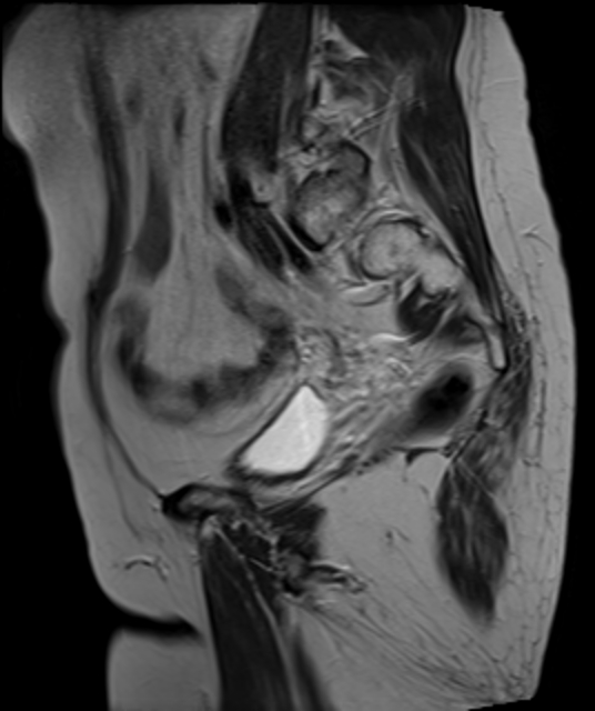
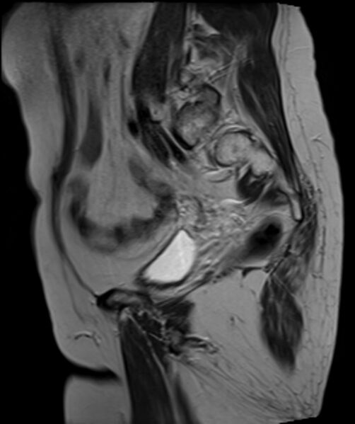
File:Cervical carcinoma (Radiopaedia 88312-104943 Sagittal T2 11).jpg
Jump to navigation
Jump to search
Size of this preview: 502 × 599 pixels. Other resolutions: 201 × 240 pixels | 536 × 640 pixels.
Original file (536 × 640 pixels, file size: 97 KB, MIME type: image/jpeg)
Summary:
| Description |
|
| Date | Published: 14th Apr 2021 |
| Source | https://radiopaedia.org/cases/cervical-carcinoma-6 |
| Author | Mehmet Yağtu |
| Permission (Permission-reusing-text) |
http://creativecommons.org/licenses/by-nc-sa/3.0/ |
Licensing:
Attribution-NonCommercial-ShareAlike 3.0 Unported (CC BY-NC-SA 3.0)
File history
Click on a date/time to view the file as it appeared at that time.
| Date/Time | Thumbnail | Dimensions | User | Comment | |
|---|---|---|---|---|---|
| current | 09:42, 2 August 2021 | | 536 × 640 (97 KB) | Fæ (talk | contribs) | Radiopaedia project rID:88312 (batch #7156-59 C11) |
You cannot overwrite this file.
File usage
There are no pages that use this file.
.jpg&oldid=865949)