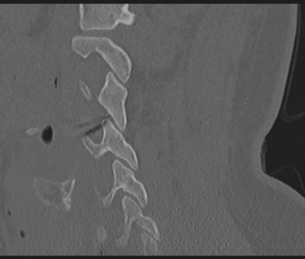
File:Cervical disc replacement (Radiopaedia 44025-47541 Sagittal bone window 57).png
Jump to navigation
Jump to search
Cervical_disc_replacement_(Radiopaedia_44025-47541_Sagittal_bone_window_57).png (602 × 512 pixels, file size: 226 KB, MIME type: image/png)
Summary:
| Description |
|
| Date | Published: 5th Apr 2016 |
| Source | https://radiopaedia.org/cases/cervical-disc-replacement-1 |
| Author | Bruno Di Muzio |
| Permission (Permission-reusing-text) |
http://creativecommons.org/licenses/by-nc-sa/3.0/ |
Licensing:
Attribution-NonCommercial-ShareAlike 3.0 Unported (CC BY-NC-SA 3.0)
File history
Click on a date/time to view the file as it appeared at that time.
| Date/Time | Thumbnail | Dimensions | User | Comment | |
|---|---|---|---|---|---|
| current | 17:12, 2 August 2021 |  | 602 × 512 (226 KB) | Fæ (talk | contribs) | Radiopaedia project rID:44025 (batch #7177-57 A57) |
You cannot overwrite this file.
File usage
The following page uses this file:
