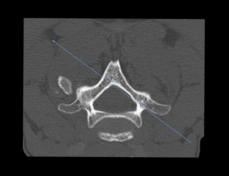
File:Cervical interlaminar epidural steroid injection (fluoroscopic guided) (Radiopaedia 77352-89457 Axial bone window 1).jpg
Jump to navigation
Jump to search

Size of this preview: 782 × 600 pixels. Other resolutions: 313 × 240 pixels | 626 × 480 pixels | 1,002 × 768 pixels | 1,280 × 981 pixels | 1,646 × 1,262 pixels.
Original file (1,646 × 1,262 pixels, file size: 198 KB, MIME type: image/jpeg)
Summary:
| Description |
|
| Date | Published: 16th May 2020 |
| Source | https://radiopaedia.org/cases/cervical-interlaminar-epidural-steroid-injection-fluoroscopic-guided |
| Author | Dai Roberts |
| Permission (Permission-reusing-text) |
http://creativecommons.org/licenses/by-nc-sa/3.0/ |
Licensing:
Attribution-NonCommercial-ShareAlike 3.0 Unported (CC BY-NC-SA 3.0)
File history
Click on a date/time to view the file as it appeared at that time.
| Date/Time | Thumbnail | Dimensions | User | Comment | |
|---|---|---|---|---|---|
| current | 23:30, 2 August 2021 |  | 1,646 × 1,262 (198 KB) | Fæ (talk | contribs) | Radiopaedia project rID:77352 (batch #7198-1 A1) |
You cannot overwrite this file.
File usage
There are no pages that use this file.