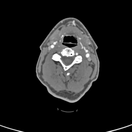
File:Cervical spine fractures with vertebral artery dissection (Radiopaedia 53296-59269 A 55).png
Jump to navigation
Jump to search
Cervical_spine_fractures_with_vertebral_artery_dissection_(Radiopaedia_53296-59269_A_55).png (512 × 512 pixels, file size: 33 KB, MIME type: image/png)
Summary:
| Description |
|
| Date | Published: 11th May 2017 |
| Source | https://radiopaedia.org/cases/cervical-spine-fractures-with-vertebral-artery-dissection-1 |
| Author | Henry Knipe |
| Permission (Permission-reusing-text) |
http://creativecommons.org/licenses/by-nc-sa/3.0/ |
Licensing:
Attribution-NonCommercial-ShareAlike 3.0 Unported (CC BY-NC-SA 3.0)
File history
Click on a date/time to view the file as it appeared at that time.
| Date/Time | Thumbnail | Dimensions | User | Comment | |
|---|---|---|---|---|---|
| current | 22:40, 3 August 2021 |  | 512 × 512 (33 KB) | Fæ (talk | contribs) | Radiopaedia project rID:53296 (batch #7261-55 A55) |
You cannot overwrite this file.
File usage
The following page uses this file:
