File:Cervical thymic cyst (Radiopaedia 88632-105336 Coronal STIR 20).jpg

From NC Commons
Jump to navigation Jump to search

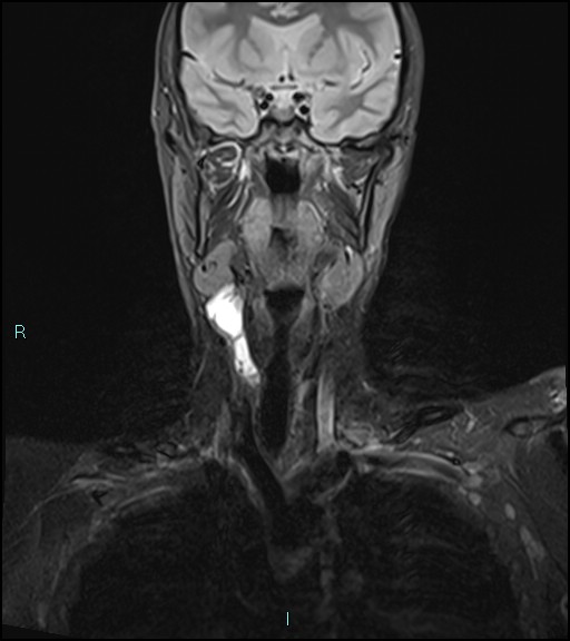
Cervical_thymic_cyst_(Radiopaedia_88632-105336_Coronal_STIR_20).jpg(512 × 576 pixels, file size: 45 KB, MIME type: image/jpeg)

Summary:

Description
  • Radiopaedia case ID: 88632
  • Image ID: 54829265
  • Image stack position: 20/38
  • Plane projection: Coronal
  • Aux modality: STIR
  • Study description: MRI neck with contrast
  • Study findings: MR imaging reveals a peripherally enhancing multi-septated cystic lesion arising in the right side of the neck (5.5 cm in total length, and 2 x 1.5 cm in the AP and transverse dimensions), with homogenous high signals on T2-weighted and low signal on T1 weighted images. The lesion lies deep to the sternocleidomastoid muscles and anterior to the carotid sheaths. No additional enhancing lesions were noted.
  • Modality: MRI
  • System: Head & Neck
  • Findings: CT Scan of the neck showed a multiloculated fluid density lesion with enhancing septae and tiny calcification in the right parapharyngeal space, extending from below the level of mandible up to C7 vertebral level. The mass was extending posteromedially to the right sternocleidomastoid muscle extending from the level of the thyroid cartilage up to the level of the superior mediastinum. There are no suspected adenopathies or recurrence of papillary thyroid cancer.
Date Published: 17th Apr 2021
Source https://radiopaedia.org/cases/cervical-thymic-cyst-1
Author Domenico Nicoletti
Permission
(Permission-reusing-text)
http://creativecommons.org/licenses/by-nc-sa/3.0/

Licensing:

Attribution-NonCommercial-ShareAlike 3.0 Unported (CC BY-NC-SA 3.0)

File history

Click on a date/time to view the file as it appeared at that time.

Date/TimeThumbnailDimensionsUserComment
current04:45, 4 August 2021Thumbnail for version as of 04:45, 4 August 2021512 × 576 (45 KB) (talk | contribs)Radiopaedia project rID:88632 (batch #7279-20 A20)

There are no pages that use this file.
Metadata