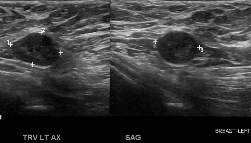
File:Changing left mammogram (Radiopaedia 23840-24001 A 1).png
Jump to navigation
Jump to search

Size of this preview: 800 × 456 pixels. Other resolutions: 320 × 183 pixels | 640 × 365 pixels | 973 × 555 pixels.
Original file (973 × 555 pixels, file size: 385 KB, MIME type: image/png)
Summary:
| Description |
|
| Date | Published: 12th Jul 2013 |
| Source | https://radiopaedia.org/cases/changing-left-mammogram |
| Author | Garth Kruger |
| Permission (Permission-reusing-text) |
http://creativecommons.org/licenses/by-nc-sa/3.0/ |
Licensing:
Attribution-NonCommercial-ShareAlike 3.0 Unported (CC BY-NC-SA 3.0)
File history
Click on a date/time to view the file as it appeared at that time.
| Date/Time | Thumbnail | Dimensions | User | Comment | |
|---|---|---|---|---|---|
| current | 01:44, 5 August 2021 | | 973 × 555 (385 KB) | Fæ (talk | contribs) | Radiopaedia project rID:23840 (batch #7323-1 A1) |
You cannot overwrite this file.
File usage
There are no pages that use this file.
.png&oldid=903011)