File:Charcot shoulder (Radiopaedia 23548-23649 Coronal T2 fat sat 15).png
Jump to navigation
Jump to search
No higher resolution available.
Charcot_shoulder_(Radiopaedia_23548-23649_Coronal_T2_fat_sat_15).png (512 × 512 pixels, file size: 100 KB, MIME type: image/png)
Summary:
| Description |
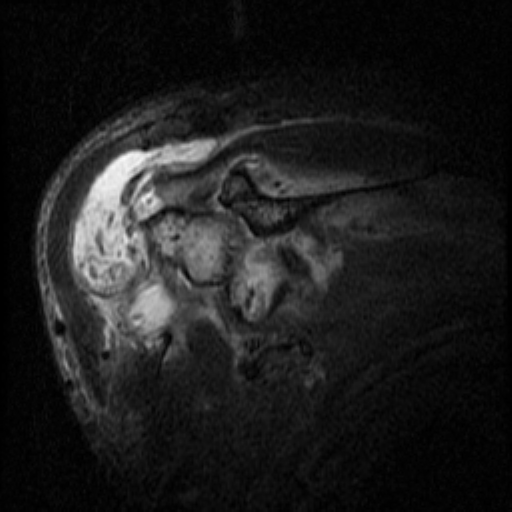
mimicking Hill-Sach's and Bankart's lesions were noted. In isolation, possibility of recurrent dislocation with septic arthritis was also considered. However, patient did not give any history of shoulder dislocation. Such a massive destruction strongly pointed towards neuropathic joint. And thus, MRI spine was done as additional study to rule out syringomyelia. |
| Date | Published: 23rd Jun 2013 |
| Source | https://radiopaedia.org/cases/charcot-shoulder |
| Author | Praveen Jha |
| Permission (Permission-reusing-text) |
http://creativecommons.org/licenses/by-nc-sa/3.0/ |
Licensing:
Attribution-NonCommercial-ShareAlike 3.0 Unported (CC BY-NC-SA 3.0)
File history
Click on a date/time to view the file as it appeared at that time.
| Date/Time | Thumbnail | Dimensions | User | Comment | |
|---|---|---|---|---|---|
| current | 14:39, 5 August 2021 | | 512 × 512 (100 KB) | Fæ (talk | contribs) | Radiopaedia project rID:23548 (batch #7356-83 E15) |
You cannot overwrite this file.
File usage
There are no pages that use this file.
.png&oldid=905925)