File:Chiari 1 - pre and post decompression surgery (Radiopaedia 42945-46188 Sagittal 31).png
Jump to navigation
Jump to search
No higher resolution available.
Chiari_1_-_pre_and_post_decompression_surgery_(Radiopaedia_42945-46188_Sagittal_31).png (515 × 512 pixels, file size: 197 KB, MIME type: image/png)
Summary:
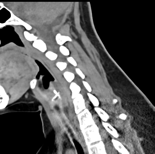
| Description |
|
| Date | Published: 27th Feb 2016 |
| Source | https://radiopaedia.org/cases/chiari-1-pre-and-post-decompression-surgery |
| Author | Bruno Di Muzio |
| Permission (Permission-reusing-text) |
http://creativecommons.org/licenses/by-nc-sa/3.0/ |
Licensing:
Attribution-NonCommercial-ShareAlike 3.0 Unported (CC BY-NC-SA 3.0)
File history
Click on a date/time to view the file as it appeared at that time.
| Date/Time | Thumbnail | Dimensions | User | Comment | |
|---|---|---|---|---|---|
| current | 11:49, 8 August 2021 | | 515 × 512 (197 KB) | Fæ (talk | contribs) | Radiopaedia project rID:42945 (batch #7429-211 F31) |
You cannot overwrite this file.
File usage
The following page uses this file:
.png&oldid=929545)