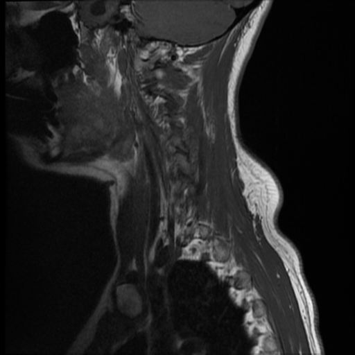
File:Chiari I malformation and obstructive hydrocephalus (Radiopaedia 41185-43984 Sagittal T1 12).png

From NC Commons
Jump to navigation Jump to search

Chiari_I_malformation_and_obstructive_hydrocephalus_(Radiopaedia_41185-43984_Sagittal_T1_12).png(512 × 512 pixels, file size: 225 KB, MIME type: image/png)

Summary:

Description
  • Radiopaedia case ID: 41185
  • Image ID: 17531612
  • Image stack position: 12/12
  • Plane projection: Sagittal
  • Aux modality: T1
  • Study description: MRI Cervical spine
  • Study findings: Standard T1 and T2 weighted sagittal and axial scans were performed through the cervical spine. A typical Chiari I malformation is noted at the foramen magnum. The cerebellar tonsils extend inferiorly as far as the superior aspect of the arch of C2. The combination of the ectopic tonsils and the medulla obliterate almost all the subarachnoid space at the foramen magnum level. The dorsal columns are convex posteriorly at the proximal end of the cervical cord. The obex is prominent, but the central canal quickly resumes normal diameter and there is no evidence of a syrinx. Incidental hemangioma noted in the body of C6. Enlarged, "empty" sella also noted. Conclusion: Chiari I malformation, but no syrinx.
  • Modality: MRI
  • System: Central Nervous System
  • Findings: Pre-and post-contrast scans of the brain including dedicated pituitary fossa imaging were performed. There is evidence of a Chiari malformation with elongated cerebellar tonsils extending 2 cm below the foramen magnum. There is marked crowding at foramen magnum with mass effect on the upper cervical cord and brainstem. No evidence of syrinx within the upper cervical cord. There is secondary obstructive hydrocephalus with marked lateral and third ventriculomegaly. The floor of the third ventricle is bowed into the suprasellar region. No evidence of transependymal edema, however, the cerebral cortical sulci appear mildly effaced. The sella appears "empty" with a prominent suprasellar cistern and pituitary tissue compressed along the floor of the sella. No pituitary mass lesion is identified. Impression: Chiari one malformation with obstructive hydrocephalus. No MRI evidence of a pituitary mass lesion.
Date Published: 22nd Nov 2015
Source https://radiopaedia.org/cases/chiari-i-malformation-and-obstructive-hydrocephalus
Author Rajalakshmi Ramesh
Permission
(Permission-reusing-text)
http://creativecommons.org/licenses/by-nc-sa/3.0/

Licensing:

Attribution-NonCommercial-ShareAlike 3.0 Unported (CC BY-NC-SA 3.0)

File history

Click on a date/time to view the file as it appeared at that time.

Date/TimeThumbnailDimensionsUserComment
current01:10, 9 August 2021Thumbnail for version as of 01:10, 9 August 2021512 × 512 (225 KB) (talk | contribs)Radiopaedia project rID:41185 (batch #7464-12 A12)

There are no pages that use this file.
Metadata