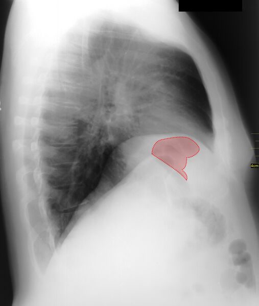
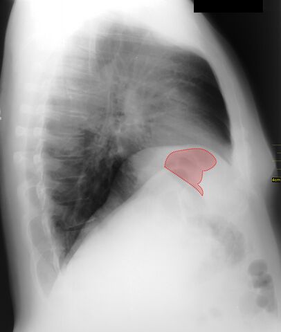
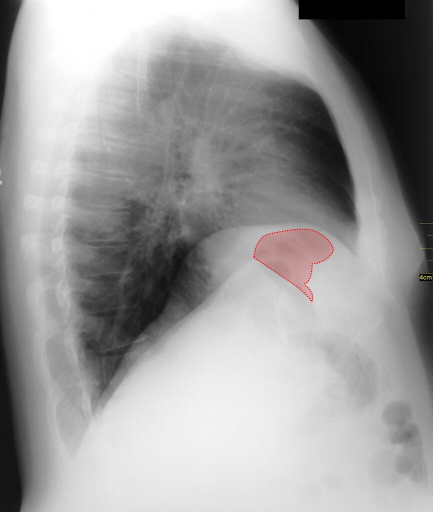
File:Chilaiditi syndrome (Radiopaedia 21936-21930 D 1).jpg
Jump to navigation
Jump to search

Size of this preview: 507 × 599 pixels. Other resolutions: 203 × 240 pixels | 406 × 480 pixels | 649 × 768 pixels | 866 × 1,024 pixels | 2,412 × 2,852 pixels.
Original file (2,412 × 2,852 pixels, file size: 2.19 MB, MIME type: image/jpeg)
Summary:
| Description |
|
| Date | 27 Feb 2013 |
| Source | Chilaiditi syndrome |
| Author | Matt Skalski |
| Permission (Permission-reusing-text) |
http://creativecommons.org/licenses/by-nc-sa/3.0/ |
Licensing:
Attribution-NonCommercial-ShareAlike 3.0 Unported (CC BY-NC-SA 3.0)
| This file is ineligible for copyright and therefore in the public domain, because it is a technical image created as part of a standard medical diagnostic procedure. No creative element rising above the threshold of originality was involved in its production.
|
 |
File history
Click on a date/time to view the file as it appeared at that time.
| Date/Time | Thumbnail | Dimensions | User | Comment | |
|---|---|---|---|---|---|
| current | 16:35, 9 August 2021 | | 2,412 × 2,852 (2.19 MB) | Fæ (talk | contribs) | Radiopaedia project rID:21936 (batch #7515-4 D1) |
You cannot overwrite this file.
File usage
There are no pages that use this file.
.jpg&oldid=9614998)