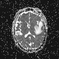
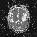
File:Chloroma - acute myeloid leukemia (Radiopaedia 66668-75972 Axial ADC 32).jpg

From NC Commons
Jump to navigation Jump to search

Chloroma_-_acute_myeloid_leukemia_(Radiopaedia_66668-75972_Axial_ADC_32).jpg(128 × 128 pixels, file size: 9 KB, MIME type: image/jpeg)

Summary:

Description
  • Radiopaedia case ID: 66668
  • Image ID: 47124352
  • Image stack position: 32/63
  • Plane projection: Axial
  • Aux modality: ADC
  • Study findings: 5.8 (AP) x 3.4 (Tran) x 2.9 (cc) cm rim-enhancing mass within the anterior inferior left temporal fossa which appears to have a dural tail extending along the surface of Meckel's cave, suggesting that this is an extra-axial dural-based mass with probable intra-axial extension in the left temporal lobe. The mass demonstrates heterogenous predominantly isointense signal on T2 weighted imaging and is mildly hypointense on T1 weighted imaging. There is some reduced diffusion within the within the solidly enhancing components of the mass, compatible with hypercellular tumor. Transosseous extension of enhancement into the infratemporal fossa and into the deep aspect of the temporalis muscle with bony changes better seen on the CT. Scattered punctate foci of susceptibility artifact consistent with hemorrhage. Associated vasogenic edema and mass effect with deviation of the left middle cerebral artery anteriorly, minimal effacement of the left lateral ventricle and unchanged minimal midline shift to the right by approximately 5 mm with slight compression of the left cerebral peduncle and minimal left uncal herniation.
  • Modality: MRI
  • System: Central Nervous System
  • Findings: 4.8 cm x 3.2 cm (axial) x 2.9 cm (craniocaudal) heterogenous mass centered in the anterior left temporal lobe. The crescentic peripheral portion of the lesion demonstrates hyperattenuation relative to the remainder of the brain parenchyma on noncontrast study and demonstrates avid enhancement on post-contrast imaging. Low attenuation centrally in the lesion consistent with necrosis. Surrounding vasogenic edema, most pronounced in the left temporal and parietal lobes with associated mass effect, with effacement of the sulcal pattern of the left temporal and parietal lobes, slight effacement of the left basal ganglia, basal cisterns, and of the left lateral ventricle; with minimal (2 mm) left-to-right midline shift. Sclerotic changes noted in the anterior left temporal bone with evidence of transosseous extension of tumor better seen on the MRI.
Date Published: 1st Mar 2019
Source https://radiopaedia.org/cases/chloroma-acute-myeloid-leukaemia
Author Danielle Byrne
Permission
(Permission-reusing-text)
http://creativecommons.org/licenses/by-nc-sa/3.0/

Licensing:

Attribution-NonCommercial-ShareAlike 3.0 Unported (CC BY-NC-SA 3.0)

File history

Click on a date/time to view the file as it appeared at that time.

Date/TimeThumbnailDimensionsUserComment
current18:08, 9 August 2021Thumbnail for version as of 18:08, 9 August 2021128 × 128 (9 KB) (talk | contribs)Radiopaedia project rID:66668 (batch #7526-226 H32)