File:Cholangiocarcinoma (Radiopaedia 49009-54379 A 151).jpg

From NC Commons
Jump to navigation Jump to search

Cholangiocarcinoma_(Radiopaedia_49009-54379_A_151).jpg(584 × 584 pixels, file size: 108 KB, MIME type: image/jpeg)

Summary:

Description
  • Radiopaedia case ID: 49009
  • Image ID: 26861304
  • Image stack position: 151/243
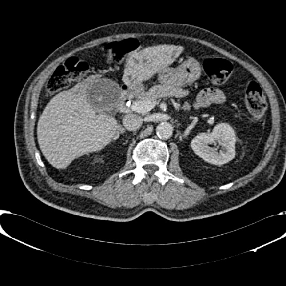
  • Plane projection: Axial
  • Aux modality: C+ portal venous phase
  • Study description: CT performed as part of staging study
  • Study findings: CT ABDOMEN CBD stent is in situ, extending into right hepatic duct. Moderate dilatation of intrahepatic biliary ducts in the left hepatic lobe. Ill defined mass measuring 4.3 x 5.4 cm in segment 4. Small volume periportal lymph nodes, measuring upto 8mm. No significant para-aortic lymphadenopathy. No ascites. The gallbladder is distended and contains stones. Spleen, pancreas, both kidneys and adrenals are unremarkable.
  • Modality: CT
  • System: Hepatobiliary
  • Findings: MRI LIVER Severe diffuse intrahepatic duct dilatation down to the porta hepatis, with an abrupt cut off with a common hepatic and bile duct of normal caliber. Approximately 6cm ill defined mass in segment 4a/b extending to the porta, at the site of the aforementioned cut-off. No synchronous hepatic lesions. Gallbladder packed with stones.  Normal wall thickness. The remainder of the solid organs are normal. No portal nodes.
Date Published: 12th Dec 2016
Source https://radiopaedia.org/cases/cholangiocarcinoma-6
Author Ian Bickle
Permission
(Permission-reusing-text)
http://creativecommons.org/licenses/by-nc-sa/3.0/

Licensing:

Attribution-NonCommercial-ShareAlike 3.0 Unported (CC BY-NC-SA 3.0)

File history

Click on a date/time to view the file as it appeared at that time.

Date/TimeThumbnailDimensionsUserComment
current02:02, 10 August 2021Thumbnail for version as of 02:02, 10 August 2021584 × 584 (108 KB) (talk | contribs)Radiopaedia project rID:49009 (batch #7546-151 A151)

The following page uses this file: