File:Cholangiocarcinoma (Radiopaedia 85717-101511 F 72).jpg

From NC Commons
Jump to navigation Jump to search

Cholangiocarcinoma_(Radiopaedia_85717-101511_F_72).jpg(508 × 588 pixels, file size: 34 KB, MIME type: image/jpeg)

Summary:

Description
  • Radiopaedia case ID: 85717
  • Image ID: 54184376
  • Image stack position: 72/96
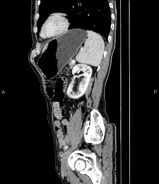
  • Plane projection: Sagittal
  • Aux modality: C+ portal venous phase
  • Study findings: Parenchymal atrophic changes and capsular retraction are seen at anterolateral aspect of liver particularly segments IV, V and VIII. The gallbladder is contracted. Intrahepatic bile ducts are dilated without dilatation of extrahepatic bile ducts. An ill defined mass like lesion is observed at connection site of right and left intrahepatic bile ducts.A few non-enhanced simple cortical cysts are seen at both kidneys.The prostate gland is enlarged.Degenerative changes as osteophytosis are seen at the thoracolumbar spine.
  • Modality: CT
  • System: Oncology
  • Findings: Mild to moderate dilatation is seen at intrahepatic bile ducts. There is no normal connection between right and left intrahepatic bile ducts. Common hepatic and common bile ducts are not dilated. Additionally parenchymal atrophic changes are noted at 4th hepatic segment.Gallbladder is contracted.
Date Published: 29th Jan 2021
Source https://radiopaedia.org/cases/cholangiocarcinoma-18
Author Mohammad Taghi Niknejad
Permission
(Permission-reusing-text)
http://creativecommons.org/licenses/by-nc-sa/3.0/

Licensing:

Attribution-NonCommercial-ShareAlike 3.0 Unported (CC BY-NC-SA 3.0)

File history

Click on a date/time to view the file as it appeared at that time.

Date/TimeThumbnailDimensionsUserComment
current05:25, 10 August 2021Thumbnail for version as of 05:25, 10 August 2021508 × 588 (34 KB) (talk | contribs)Radiopaedia project rID:85717 (batch #7548-501 F72)

The following page uses this file: