File:Cholangiocarcinoma - hilar type (Radiopaedia 59780-67206 M 43).jpg

From NC Commons
Jump to navigation Jump to search

Cholangiocarcinoma_-_hilar_type_(Radiopaedia_59780-67206_M_43).jpg(320 × 260 pixels, file size: 24 KB, MIME type: image/jpeg)

Summary:

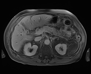
Description
  • Radiopaedia case ID: 59780
  • Image ID: 37997261
  • Image stack position: 43/72
  • Plane projection: Axial
  • Aux modality: T1 C+ fat sat
  • Study findings: A hilar mass measuring 15 x 25 mm (transverse) is identified, resulting in marked intrahepatic duct dilatation. The stricture extends over a length of approximately 15 mm.Gradual enhancement of the lesion is demonstrated on the dynamic series with diffusion restriction present. Beaking of the proximal left intra-hepatic duct with separation of the right anterior and posterior sectoral ducts. No liver metastases. The common bile duct is normal.  No periportal or retroperitoneal lymphadenopathy. Portal vein and right and left branches are patent.  Hepatic veins are patent.Gallbladder decompressed.
  • Modality: MRI
  • System: Hepatobiliary
  • Findings: There is extensive intrahepatic duct dilatation, more pronounced in the left hepatic lobe. The common hepatic duct shows diffusely thick and enhancing walls. The gallbladder is contracted and shows an irregular enhancing mucosa. The common bile duct is difficult to visualize and hence not dilated. A few prominent but subcentimeter celiac lymph nodes are noted. Normal enhancement of the pancreatic parenchyma on this study phase with no definite pancreatic head mass identified and the pancreatic duct is not dilated. No intra-hepatic lesion identified. The portal veins, splenic and superior mesenteric veins enhance normally.  No adenopathy elsewhere in the abdomen or pelvis. There are a small sliding-type hiatal hernia and an incidental splenunculus anterior to the spleen. The spleen, kidneys and adrenals are otherwise unremarkable. The small and large bowel appear to be within normal limits. Widespread, severe cylindrical bronchiectasis at the imaged lung bases with a large number of bronchi either containing air-fluid levels or occluded by mucus. In addition, however, there are several small peripheral nodular densities which do not appear to be related to the bronchi and so are suspicious for pulmonary metastases.
Date Published: 5th Aug 2018
Source https://radiopaedia.org/cases/cholangiocarcinoma-hilar-type
Author Bruno Di Muzio
Permission
(Permission-reusing-text)
http://creativecommons.org/licenses/by-nc-sa/3.0/

Licensing:

Attribution-NonCommercial-ShareAlike 3.0 Unported (CC BY-NC-SA 3.0)

File history

Click on a date/time to view the file as it appeared at that time.

Date/TimeThumbnailDimensionsUserComment
current08:52, 10 August 2021Thumbnail for version as of 08:52, 10 August 2021320 × 260 (24 KB) (talk | contribs)Radiopaedia project rID:59780 (batch #7551-758 M43)