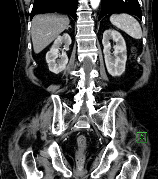
File:Cholangiocarcinoma with cerebral metastases (Radiopaedia 83519-98629 E 63).jpg
Jump to navigation
Jump to search
Size of this preview: 531 × 600 pixels. Other resolutions: 212 × 240 pixels | 632 × 714 pixels.
Original file (632 × 714 pixels, file size: 111 KB, MIME type: image/jpeg)
Summary:
| Description |
|
| Date | Published: 28th Oct 2020 |
| Source | https://radiopaedia.org/cases/cholangiocarcinoma-with-cerebral-metastases |
| Author | Ammar Ashraf |
| Permission (Permission-reusing-text) |
http://creativecommons.org/licenses/by-nc-sa/3.0/ |
Licensing:
Attribution-NonCommercial-ShareAlike 3.0 Unported (CC BY-NC-SA 3.0)
File history
Click on a date/time to view the file as it appeared at that time.
| Date/Time | Thumbnail | Dimensions | User | Comment | |
|---|---|---|---|---|---|
| current | 12:24, 10 August 2021 | | 632 × 714 (111 KB) | Fæ (talk | contribs) | Radiopaedia project rID:83519 (batch #7553-663 E63) |
You cannot overwrite this file.
File usage
The following page uses this file:
.jpg&oldid=962592)