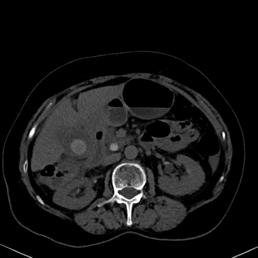
File:Cholecystitis - CT IVC obstructive choledocholitiasis (Radiopaedia 43966-47479 C 47).png
Jump to navigation
Jump to search
Cholecystitis_-_CT_IVC_obstructive_choledocholitiasis_(Radiopaedia_43966-47479_C_47).png (512 × 512 pixels, file size: 128 KB, MIME type: image/png)
Summary:
| Description |
|
| Date | Published: 2nd Apr 2016 |
| Source | https://radiopaedia.org/cases/cholecystitis-ct-ivc-obstructive-choledocholitiasis |
| Author | Bruno Di Muzio |
| Permission (Permission-reusing-text) |
http://creativecommons.org/licenses/by-nc-sa/3.0/ |
Licensing:
Attribution-NonCommercial-ShareAlike 3.0 Unported (CC BY-NC-SA 3.0)
File history
Click on a date/time to view the file as it appeared at that time.
| Date/Time | Thumbnail | Dimensions | User | Comment | |
|---|---|---|---|---|---|
| current | 14:48, 10 August 2021 |  | 512 × 512 (128 KB) | Fæ (talk | contribs) | Radiopaedia project rID:43966 (batch #7557-175 C47) |
You cannot overwrite this file.
File usage
The following page uses this file:
