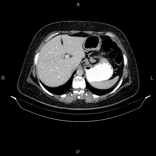
File:Choledochal cyst - type1 (Radiopaedia 83957-99176 A 23).jpg
Jump to navigation
Jump to search
Choledochal_cyst_-_type1_(Radiopaedia_83957-99176_A_23).jpg (512 × 512 pixels, file size: 32 KB, MIME type: image/jpeg)
Summary:
| Description |
|
| Date | Published: 9th Nov 2020 |
| Source | https://radiopaedia.org/cases/choledochal-cyst-type1-1 |
| Author | Mohammad Taghi Niknejad |
| Permission (Permission-reusing-text) |
http://creativecommons.org/licenses/by-nc-sa/3.0/ |
Licensing:
Attribution-NonCommercial-ShareAlike 3.0 Unported (CC BY-NC-SA 3.0)
File history
Click on a date/time to view the file as it appeared at that time.
| Date/Time | Thumbnail | Dimensions | User | Comment | |
|---|---|---|---|---|---|
| current | 19:24, 10 August 2021 |  | 512 × 512 (32 KB) | Fæ (talk | contribs) | Radiopaedia project rID:83957 (batch #7572-23 A23) |
You cannot overwrite this file.
File usage
The following file is a duplicate of this file (more details):
The following page uses this file:
