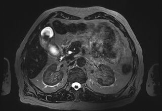
File:Choledocholithiasis (Radiopaedia 73235-83968 D 25).jpg
Jump to navigation
Jump to search

Size of this preview: 800 × 551 pixels. Other resolutions: 320 × 220 pixels | 640 × 441 pixels | 897 × 618 pixels.
Original file (897 × 618 pixels, file size: 219 KB, MIME type: image/jpeg)
Summary:
| Description |
|
| Date | Published: 4th Jan 2020 |
| Source | https://radiopaedia.org/cases/choledocholithiasis-37 |
| Author | Mostafa El-Feky |
| Permission (Permission-reusing-text) |
http://creativecommons.org/licenses/by-nc-sa/3.0/ |
Licensing:
Attribution-NonCommercial-ShareAlike 3.0 Unported (CC BY-NC-SA 3.0)
File history
Click on a date/time to view the file as it appeared at that time.
| Date/Time | Thumbnail | Dimensions | User | Comment | |
|---|---|---|---|---|---|
| current | 17:56, 17 August 2021 | | 897 × 618 (219 KB) | Fæ (talk | contribs) | Radiopaedia project rID:73235 (batch #7640-98 D25) |
You cannot overwrite this file.
File usage
The following page uses this file:
.jpg&oldid=1018947)