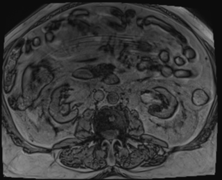
File:Choledocholithiasis and gastric polyp (Radiopaedia 48856-53896 C 63).png
Jump to navigation
Jump to search
Choledocholithiasis_and_gastric_polyp_(Radiopaedia_48856-53896_C_63).png (320 × 260 pixels, file size: 44 KB, MIME type: image/png)
Summary:
| Description |
|
| Date | Published: 7th Nov 2016 |
| Source | https://radiopaedia.org/cases/choledocholithiasis-and-gastric-polyp |
| Author | Paul Simkin |
| Permission (Permission-reusing-text) |
http://creativecommons.org/licenses/by-nc-sa/3.0/ |
Licensing:
Attribution-NonCommercial-ShareAlike 3.0 Unported (CC BY-NC-SA 3.0)
File history
Click on a date/time to view the file as it appeared at that time.
| Date/Time | Thumbnail | Dimensions | User | Comment | |
|---|---|---|---|---|---|
| current | 21:38, 11 August 2021 |  | 320 × 260 (44 KB) | Fæ (talk | contribs) | Radiopaedia project rID:48856 (batch #7636-117 C63) |
You cannot overwrite this file.
File usage
The following page uses this file:
