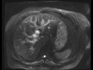
File:Choledocholithiasis causing intrahepatic biliary duct dilation (Radiopaedia 39908-42369 Axial T2 SPAIR 48).jpg
Jump to navigation
Jump to search

Size of this preview: 800 × 600 pixels. Other resolutions: 320 × 240 pixels | 640 × 480 pixels | 1,024 × 768 pixels.
Original file (1,024 × 768 pixels, file size: 179 KB, MIME type: image/jpeg)
Summary:
| Description |
|
| Date | Published: 1st Oct 2015 |
| Source | https://radiopaedia.org/cases/choledocholithiasis-causing-intrahepatic-biliary-duct-dilation |
| Author | Subash Thapa |
| Permission (Permission-reusing-text) |
http://creativecommons.org/licenses/by-nc-sa/3.0/ |
Licensing:
Attribution-NonCommercial-ShareAlike 3.0 Unported (CC BY-NC-SA 3.0)
File history
Click on a date/time to view the file as it appeared at that time.
| Date/Time | Thumbnail | Dimensions | User | Comment | |
|---|---|---|---|---|---|
| current | 23:18, 11 August 2021 | | 1,024 × 768 (179 KB) | Fæ (talk | contribs) | Radiopaedia project rID:39908 (batch #7638-68 B48) |
You cannot overwrite this file.
File usage
There are no pages that use this file.
.jpg&oldid=983806)