File:Cholesterol granuloma of the petrous apex (Radiopaedia 64358-73140 Axial TOF 3D 61).jpg
Jump to navigation
Jump to search
No higher resolution available.
Cholesterol_granuloma_of_the_petrous_apex_(Radiopaedia_64358-73140_Axial_TOF_3D_61).jpg (512 × 512 pixels, file size: 32 KB, MIME type: image/jpeg)
Summary:
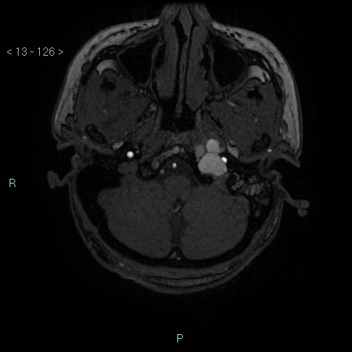
| Description |
|
| Date | Published: 19th Nov 2018 |
| Source | https://radiopaedia.org/cases/cholesterol-granuloma-of-the-petrous-apex |
| Author | Domenico Nicoletti |
| Permission (Permission-reusing-text) |
http://creativecommons.org/licenses/by-nc-sa/3.0/ |
Licensing:
Attribution-NonCommercial-ShareAlike 3.0 Unported (CC BY-NC-SA 3.0)
File history
Click on a date/time to view the file as it appeared at that time.
| Date/Time | Thumbnail | Dimensions | User | Comment | |
|---|---|---|---|---|---|
| current | 02:39, 13 August 2021 | | 512 × 512 (32 KB) | Fæ (talk | contribs) | Radiopaedia project rID:64358 (batch #7706-170 E61) |
You cannot overwrite this file.
File usage
The following page uses this file:
.jpg&oldid=997048)