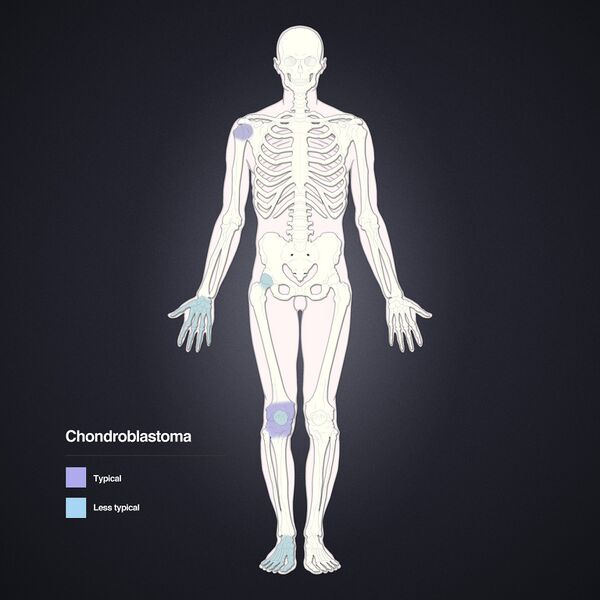
File:Chondroblastoma - distal femur (Radiopaedia 8112-15916 A 1).jpg
Jump to navigation
Jump to search

Size of this preview: 600 × 600 pixels. Other resolutions: 240 × 240 pixels | 480 × 480 pixels | 768 × 768 pixels | 1,024 × 1,024 pixels | 2,048 × 2,048 pixels.
Original file (2,048 × 2,048 pixels, file size: 1.22 MB, MIME type: image/jpeg)
Summary:
| Description |
|
| Date | Published: 13th Jan 2010 |
| Source | https://radiopaedia.org/cases/chondroblastoma-distal-femur-2 |
| Author | Angela Byrne |
| Permission (Permission-reusing-text) |
http://creativecommons.org/licenses/by-nc-sa/3.0/ |
Licensing:
Attribution-NonCommercial-ShareAlike 3.0 Unported (CC BY-NC-SA 3.0)
File history
Click on a date/time to view the file as it appeared at that time.
| Date/Time | Thumbnail | Dimensions | User | Comment | |
|---|---|---|---|---|---|
| current | 05:43, 13 August 2021 | | 2,048 × 2,048 (1.22 MB) | Fæ (talk | contribs) | Radiopaedia project rID:8112 (batch #7727-1 A1) |
You cannot overwrite this file.
File usage
There are no pages that use this file.
.jpg&oldid=999213)