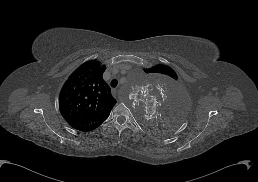
File:Chondrosarcoma - chest wall (Radiopaedia 65192-74199 Axial bone window 34).jpg

From NC Commons
Jump to navigation Jump to search

Chondrosarcoma_-_chest_wall_(Radiopaedia_65192-74199_Axial_bone_window_34).jpg(512 × 362 pixels, file size: 51 KB, MIME type: image/jpeg)

Summary:

Description
  • Radiopaedia case ID: 65192
  • Image ID: 44995140
  • Image stack position: 34/64
  • Plane projection: Axial
  • Aux modality: bone window
  • Study findings: Large left-sided chest wall mass lesion with intra and extrathoracic extension developed mainly on the left 2nd rib, but involving the left 3rd rib. The mass causes bony erosion of the T2 and to a lesser extent T3 vertebral body, the tumoral tissues extending to the left neural foramen of T2-T3 with mild extension to the epidural space without significant compression of the spinal cord. The mass is well-defined, predominantly hypodense with central calcified component. It is extending to the mediastinum, abutting the left subclavian artery, aortic arch, and the left pulmonary artery.  The mass is causing compression and narrowing of the left brachiocephalic vein although it is patent; however, the left subclavian vein is not well seen. The mass is also compressing and displacing the upper mediastinal structures, including the esophagus and trachea. Preserved fat planes between the mass and the erector spinae muscles, which are compressed on the left side, as well as the trapezius and rhomboid muscles on the left side. Mild collapse of the left lung parenchymal tissue inferior to the tumor. No suspicious lung nodules or masses. Small cervical lymph nodules are likely reactive nodes. No visualized lymphadenopathy.  The imaging appearance of the mass is suggestive of chondrosarcoma.
  • Modality: CT
  • System: Musculoskeletal
  • Findings: There is a large left upper lung zone partially defined mass with internal calcifications exerting mass effect on the trachea and the adjacent lung parenchyma and extending above the clavicle suggesting a posterior location (cervicothoracic sign). The adjacent ribs are not well visualized representing rib destruction. No airspace consolidation or lung masses. Heart is normal in size. No pleural effusion or pneumothorax seen.
Date Published: 29th Dec 2018
Source https://radiopaedia.org/cases/chondrosarcoma-chest-wall-1
Author Yasser Asiri
Permission
(Permission-reusing-text)
http://creativecommons.org/licenses/by-nc-sa/3.0/

Licensing:

Attribution-NonCommercial-ShareAlike 3.0 Unported (CC BY-NC-SA 3.0)

File history

Click on a date/time to view the file as it appeared at that time.

Date/TimeThumbnailDimensionsUserComment
current17:26, 13 August 2021Thumbnail for version as of 17:26, 13 August 2021512 × 362 (51 KB) (talk | contribs)Radiopaedia project rID:65192 (batch #7779-34 A34)