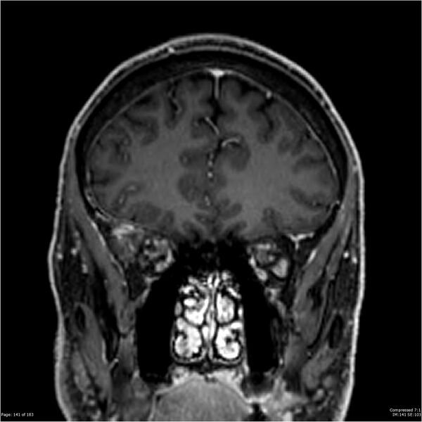
File:Chondrosarcoma of skull base- grade II (Radiopaedia 40948-43654 Coronal T1 C+ 52).jpg

From NC Commons
Jump to navigation Jump to search

Original file(2,133 × 2,133 pixels, file size: 187 KB, MIME type: image/jpeg)

Summary:

Description
  • Radiopaedia case ID: 40948
  • Image ID: 17272469
  • Image stack position: 52/60
  • Plane projection: Coronal
  • Aux modality: T1 C+
  • Study description: MRI Brain
  • Study findings: A well defined lobulated mass measuring 3.5 x 3.5 x 5.8 cm centered at the right clivus, crossing the midline by 4 mm, is heterogeneous, but predominantly T1W hypointense, brightly T2W hyperintense, with loculated/bubbly internal enhancement. It has a well-defined thin sclerotic margin medially, and an ill-defined permeative margin laterally with mass effect upon the right anterior mesiotemporal lobe. There is breach of bony cortex posteriorly with a 14 x 6 mm soft tissue projection that protrudes into the prepontine cistern, beneath dura, but lying in close proximity to the basilar artery. There is also destruction of the right carotid canal with small foci of ossific debris/coarse calcification that are T1W hyperintense. The right cavernous carotid artery is displaced, but its course lies predominantly inferomedial. Normal cavernous carotid flow void is preserved although mild narrowing is present. The right cavernous sinus and Meckel's cave are obliterated. The lateral wall of the right sphenoid sinus is remodeled, and intact, with the sinus well aerated. Part of the superolateral wall of the right foramen rotundum is breached. Right foramen ovale is slightly widened. The right hypoglossal canal is breached superiorly. Tumor lies close to the right optic canal, but the optic canal is not compromised. Inferiorly, a tongue of tissue has protruded into the right longus capitis muscles, but is 7 mm from the right nasopharyngeal mucosa. Right cochlear and vestibular apparati, middle ear and mastoid uninvolved. Right jugular foramen also appears uninvolved. Minor chronic small vessel ischemic change. No diffusion restriction. Thickened left maxillary sinus walls with mild mucosal thickening in keeping with chronic sinusitis. Conclusion: Right central skull base tumor most likely to represent chondrosarcoma or (eccentric) chordoma with extent as described.
  • Modality: MRI
  • System: Central Nervous System
  • Findings: Refer to MRI report below for combined CT and MRI image findings.
Date Published: 11th Nov 2015
Source https://radiopaedia.org/cases/chondrosarcoma-of-skull-base-grade-ii
Author Rajalakshmi Ramesh
Permission
(Permission-reusing-text)
http://creativecommons.org/licenses/by-nc-sa/3.0/

Licensing:

Attribution-NonCommercial-ShareAlike 3.0 Unported (CC BY-NC-SA 3.0)

File history

Click on a date/time to view the file as it appeared at that time.

Date/TimeThumbnailDimensionsUserComment
current02:37, 14 August 2021Thumbnail for version as of 02:37, 14 August 20212,133 × 2,133 (187 KB) (talk | contribs)Radiopaedia project rID:40948 (batch #7791-346 H52)