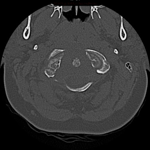
File:Chordoma - clivus (Radiopaedia 12796-12928 Axial non-contrast 7).jpg
Jump to navigation
Jump to search
No higher resolution available.
Chordoma_-_clivus_(Radiopaedia_12796-12928_Axial_non-contrast_7).jpg (512 × 512 pixels, file size: 53 KB, MIME type: image/jpeg)
Summary:
| Description |
|
| Date | Published: 11th Jan 2011 |
| Source | https://radiopaedia.org/cases/chordoma-clivus-3 |
| Author | Frank Gaillard |
| Permission (Permission-reusing-text) |
http://creativecommons.org/licenses/by-nc-sa/3.0/ |
Licensing:
Attribution-NonCommercial-ShareAlike 3.0 Unported (CC BY-NC-SA 3.0)
File history
Click on a date/time to view the file as it appeared at that time.
| Date/Time | Thumbnail | Dimensions | User | Comment | |
|---|---|---|---|---|---|
| current | 04:29, 18 August 2021 | | 512 × 512 (53 KB) | Fæ (talk | contribs) | Radiopaedia project rID:12796 (batch #7848-18 C7) |
You cannot overwrite this file.
File usage
There are no pages that use this file.
.jpg&oldid=1020803)