File:Choriocarcinoma (Radiopaedia 31889-32839 A 26).jpg
Jump to navigation
Jump to search
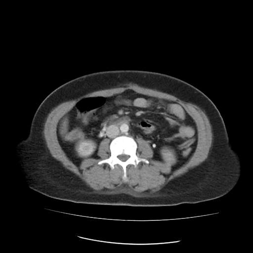
Choriocarcinoma_(Radiopaedia_31889-32839_A_26).jpg (512 × 512 pixels, file size: 18 KB, MIME type: image/jpeg)
Summary:
| Description |
|
| Date | Published: 3rd Nov 2014 |
| Source | https://radiopaedia.org/cases/choriocarcinoma-1 |
| Author | Abdel-Rahman Abdel-Halim |
| Permission (Permission-reusing-text) |
http://creativecommons.org/licenses/by-nc-sa/3.0/ |
Licensing:
Attribution-NonCommercial-ShareAlike 3.0 Unported (CC BY-NC-SA 3.0)
File history
Click on a date/time to view the file as it appeared at that time.
| Date/Time | Thumbnail | Dimensions | User | Comment | |
|---|---|---|---|---|---|
| current | 08:02, 18 August 2021 |  | 512 × 512 (18 KB) | Fæ (talk | contribs) | Radiopaedia project rID:31889 (batch #7858-26 A26) |
You cannot overwrite this file.
File usage
The following page uses this file:
