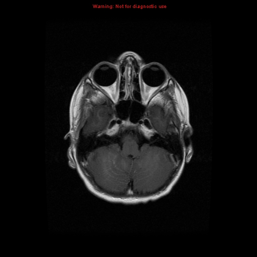
File:Choroid plexus carcinoma - recurrent (Radiopaedia 8330-9169 Axial T1 C+ 5).jpg
Jump to navigation
Jump to search
Choroid_plexus_carcinoma_-_recurrent_(Radiopaedia_8330-9169_Axial_T1_C+_5).jpg (512 × 512 pixels, file size: 69 KB, MIME type: image/jpeg)
Summary:
| Description |
|
| Date | Published: 24th Jan 2010 |
| Source | https://radiopaedia.org/cases/choroid-plexus-carcinoma-recurrent-2 |
| Author | Hani Makky Al Salam |
| Permission (Permission-reusing-text) |
http://creativecommons.org/licenses/by-nc-sa/3.0/ |
Licensing:
Attribution-NonCommercial-ShareAlike 3.0 Unported (CC BY-NC-SA 3.0)
File history
Click on a date/time to view the file as it appeared at that time.
| Date/Time | Thumbnail | Dimensions | User | Comment | |
|---|---|---|---|---|---|
| current | 17:16, 19 August 2021 |  | 512 × 512 (69 KB) | Fæ (talk | contribs) | Radiopaedia project rID:8330 (batch #7918-86 D5) |
You cannot overwrite this file.
File usage
There are no pages that use this file.
