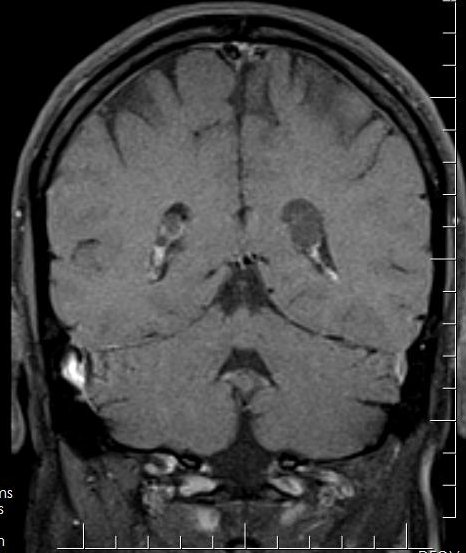
File:Choroid plexus xanthogranuloma (Radiopaedia 29624-30138 Coronal T1 1).jpg
Jump to navigation
Jump to search
Choroid_plexus_xanthogranuloma_(Radiopaedia_29624-30138_Coronal_T1_1).jpg (466 × 553 pixels, file size: 42 KB, MIME type: image/jpeg)
Summary:
| Description |
|
| Date | Published: 10th Jun 2014 |
| Source | https://radiopaedia.org/cases/choroid-plexus-xanthogranuloma-3 |
| Author | Aruna Pallewatte |
| Permission (Permission-reusing-text) |
http://creativecommons.org/licenses/by-nc-sa/3.0/ |
Licensing:
Attribution-NonCommercial-ShareAlike 3.0 Unported (CC BY-NC-SA 3.0)
File history
Click on a date/time to view the file as it appeared at that time.
| Date/Time | Thumbnail | Dimensions | User | Comment | |
|---|---|---|---|---|---|
| current | 10:13, 20 August 2021 |  | 466 × 553 (42 KB) | Fæ (talk | contribs) | Radiopaedia project rID:29624 (batch #7953-2 B1) |
You cannot overwrite this file.
File usage
There are no pages that use this file.
