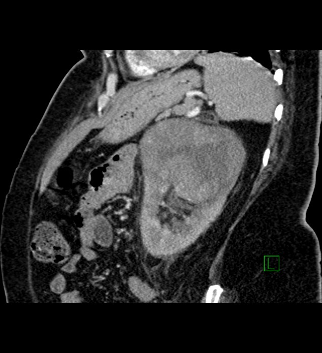
File:Chromophobe renal cell carcinoma (Radiopaedia 84239-99519 D 35).jpg
Jump to navigation
Jump to search
Size of this preview: 547 × 600 pixels. Other resolutions: 219 × 240 pixels | 634 × 695 pixels.
Original file (634 × 695 pixels, file size: 79 KB, MIME type: image/jpeg)
Summary:
| Description |
|
| Date | Published: 18th Nov 2020 |
| Source | https://radiopaedia.org/cases/chromophobe-renal-cell-carcinoma |
| Author | Ammar Ashraf |
| Permission (Permission-reusing-text) |
http://creativecommons.org/licenses/by-nc-sa/3.0/ |
Licensing:
Attribution-NonCommercial-ShareAlike 3.0 Unported (CC BY-NC-SA 3.0)
File history
Click on a date/time to view the file as it appeared at that time.
| Date/Time | Thumbnail | Dimensions | User | Comment | |
|---|---|---|---|---|---|
| current | 13:21, 20 August 2021 | | 634 × 695 (79 KB) | Fæ (talk | contribs) | Radiopaedia project rID:84239 (batch #7971-294 D35) |
You cannot overwrite this file.
File usage
The following page uses this file:
.jpg&oldid=1043535)