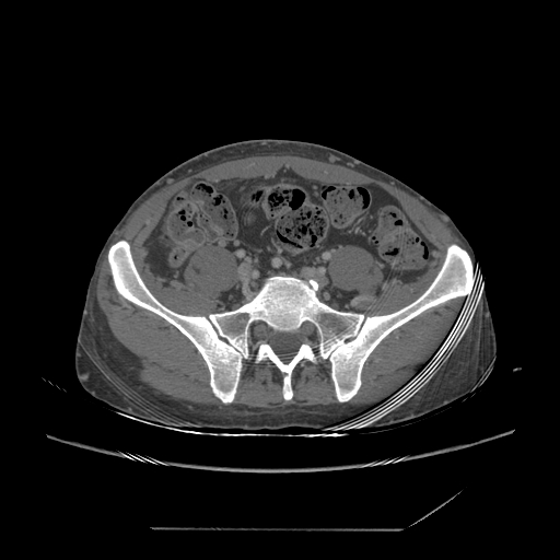
File:Chronic IVC thrombosis and resultant IVC filter malposition (Radiopaedia 81158-94800 B 63).jpg
Jump to navigation
Jump to search
Chronic_IVC_thrombosis_and_resultant_IVC_filter_malposition_(Radiopaedia_81158-94800_B_63).jpg (512 × 512 pixels, file size: 78 KB, MIME type: image/jpeg)
Summary:
| Description |
|
| Date | Published: 21st Aug 2020 |
| Source | https://radiopaedia.org/cases/chronic-ivc-thrombosis-and-resultant-ivc-filter-malposition |
| Author | Faeze Salahshour |
| Permission (Permission-reusing-text) |
http://creativecommons.org/licenses/by-nc-sa/3.0/ |
Licensing:
Attribution-NonCommercial-ShareAlike 3.0 Unported (CC BY-NC-SA 3.0)
File history
Click on a date/time to view the file as it appeared at that time.
| Date/Time | Thumbnail | Dimensions | User | Comment | |
|---|---|---|---|---|---|
| current | 01:18, 22 August 2021 |  | 512 × 512 (78 KB) | Fæ (talk | contribs) | Radiopaedia project rID:81158 (batch #8049-196 B63) |
You cannot overwrite this file.
File usage
The following page uses this file:
