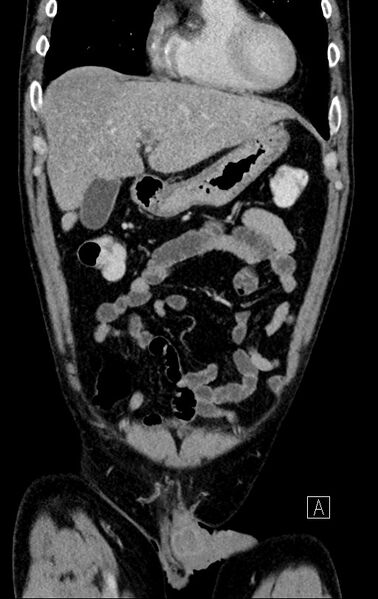
File:Chronic appendicitis complicated by appendicular abscess, pylephlebitis and liver abscess (Radiopaedia 54483-60700 C 22).jpg

From NC Commons
Jump to navigation Jump to search

Original file(512 × 811 pixels, file size: 124 KB, MIME type: image/jpeg)

Summary:

Description
  • Radiopaedia case ID: 54483
  • Image ID: 31403000
  • Image stack position: 22/79
  • Plane projection: Coronal
  • Aux modality: C+ portal venous phase
  • Study description: CT Abdomen and Pelvis
  • Modality: CT
  • System: Gastrointestinal
  • Findings: Two-phase contrast CT shows occlusion of the left main portal vein and its tributaries and subsequent hyperperfusion of the left liver lobe in the arterial phase, most likely due to consecutive predominant arterial supply of the left liver parenchyma via the hepatic artery. Evidence of a heterogeneous subcapsular hepatic lesion in segment II, measuring 4,5 x 2,7 x 3 cm. The lesion demonstrates a hypodense center and a double ring, the thin inner ring being hyperdense and the thicker outer ring hypodense, compared to the adjacent liver parenchyma. The appendix appears mildly swollen with moderate surrounding fat stranding. A fluid collection is seen medial to the tip of the appendix with a thick hyperdense wall and hypodense content alongside some gas bubbles. This formation measures 6,5 x 2 x 3 cm and abutts the urinary bladder. The right ventral bladder wall appears markedly thickened. Evidence of para-aortal lymphadenopathy. No other acute findings. Secondary findings include hepatic steatosis, small liver cysts, advanced arteriosclerosis, reperfusion of the umbilical vein and some bilateral dystelecatic changes of the dorso-basal lung parenchyma CONCLUSION: Clinical history, examination, lab results and CT features are in keeping with chronic appendicitis with appendicular abscess and a pyogenic spread of the intraabdominal infection via the portal venous drainage way. Consecutive pylephlebitis of the left portal vein with a superimposed hepatic abscess in the affected left liver segment II. The abscess demonstrates a double target sign which is a characteristic image feature of a hepatic abscess.
Date Published: 13th Jul 2017
Source https://radiopaedia.org/cases/chronic-appendicitis-complicated-by-appendicular-abscess-pylephlebitis-and-liver-abscess-1
Author Benedikt Beilstein
Permission
(Permission-reusing-text)
http://creativecommons.org/licenses/by-nc-sa/3.0/

Licensing:

Attribution-NonCommercial-ShareAlike 3.0 Unported (CC BY-NC-SA 3.0)

File history

Click on a date/time to view the file as it appeared at that time.

Date/TimeThumbnailDimensionsUserComment
current02:17, 21 August 2021Thumbnail for version as of 02:17, 21 August 2021512 × 811 (124 KB) (talk | contribs)Radiopaedia project rID:54483 (batch #7986-261 C22)