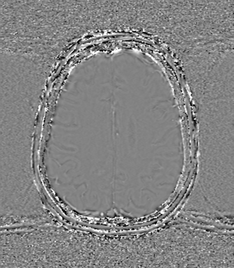
File:Chronic hypertensive encephalopathy (Radiopaedia 72844-83495 Axial SWI phase 62).png
Jump to navigation
Jump to search
Chronic_hypertensive_encephalopathy_(Radiopaedia_72844-83495_Axial_SWI_phase_62).png (336 × 384 pixels, file size: 155 KB, MIME type: image/png)
Summary:
| Description |
|
| Date | Published: 6th Apr 2020 |
| Source | https://radiopaedia.org/cases/chronic-hypertensive-encephalopathy-4 |
| Author | Frank Gaillard |
| Permission (Permission-reusing-text) |
http://creativecommons.org/licenses/by-nc-sa/3.0/ |
Licensing:
Attribution-NonCommercial-ShareAlike 3.0 Unported (CC BY-NC-SA 3.0)
File history
Click on a date/time to view the file as it appeared at that time.
| Date/Time | Thumbnail | Dimensions | User | Comment | |
|---|---|---|---|---|---|
| current | 17:45, 21 August 2021 |  | 336 × 384 (155 KB) | Fæ (talk | contribs) | Radiopaedia project rID:72844 (batch #8039-341 G62) |
You cannot overwrite this file.
File usage
The following page uses this file:
