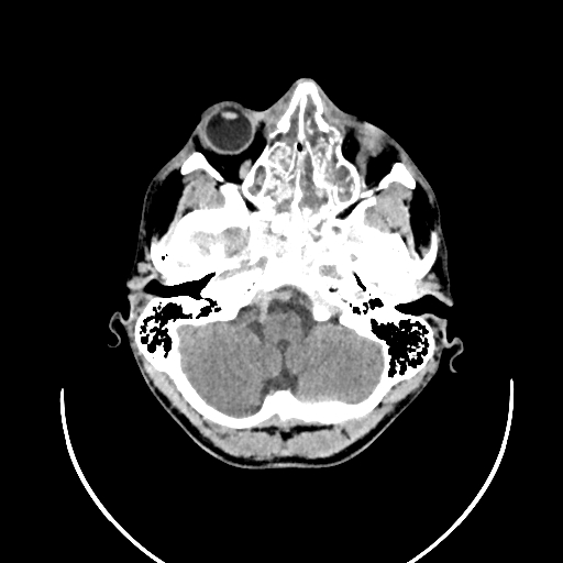
File:Chronic invasive fungal sinusitis (Radiopaedia 50342-55710 Axial non-contrast 35).jpg
Jump to navigation
Jump to search
Chronic_invasive_fungal_sinusitis_(Radiopaedia_50342-55710_Axial_non-contrast_35).jpg (512 × 512 pixels, file size: 79 KB, MIME type: image/jpeg)
Summary:
| Description |
|
| Date | Published: 5th Jan 2017 |
| Source | https://radiopaedia.org/cases/chronic-invasive-fungal-sinusitis |
| Author | Dr. Ekaterina Peshevich |
| Permission (Permission-reusing-text) |
http://creativecommons.org/licenses/by-nc-sa/3.0/ |
Licensing:
Attribution-NonCommercial-ShareAlike 3.0 Unported (CC BY-NC-SA 3.0)
File history
Click on a date/time to view the file as it appeared at that time.
| Date/Time | Thumbnail | Dimensions | User | Comment | |
|---|---|---|---|---|---|
| current | 20:40, 21 August 2021 |  | 512 × 512 (79 KB) | Fæ (talk | contribs) | Radiopaedia project rID:50342 (batch #8044-35 A35) |
You cannot overwrite this file.
File usage
The following page uses this file:
