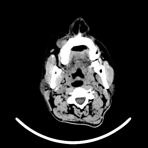
File:Chronic invasive fungal sinusitis with intraorbital and intracranial extension (Radiopaedia 56387-63046 Axial non-contrast 11).jpg
Jump to navigation
Jump to search
Chronic_invasive_fungal_sinusitis_with_intraorbital_and_intracranial_extension_(Radiopaedia_56387-63046_Axial_non-contrast_11).jpg (512 × 512 pixels, file size: 64 KB, MIME type: image/jpeg)
Summary:
| Description |
|
| Date | Published: 29th Oct 2017 |
| Source | https://radiopaedia.org/cases/chronic-invasive-fungal-sinusitis-with-intraorbital-and-intracranial-extension |
| Author | Mohammad Farghali Ali Tosson |
| Permission (Permission-reusing-text) |
http://creativecommons.org/licenses/by-nc-sa/3.0/ |
Licensing:
Attribution-NonCommercial-ShareAlike 3.0 Unported (CC BY-NC-SA 3.0)
File history
Click on a date/time to view the file as it appeared at that time.
| Date/Time | Thumbnail | Dimensions | User | Comment | |
|---|---|---|---|---|---|
| current | 21:38, 21 August 2021 |  | 512 × 512 (64 KB) | Fæ (talk | contribs) | Radiopaedia project rID:56387 (batch #8045-11 A11) |
You cannot overwrite this file.
File usage
The following page uses this file:
