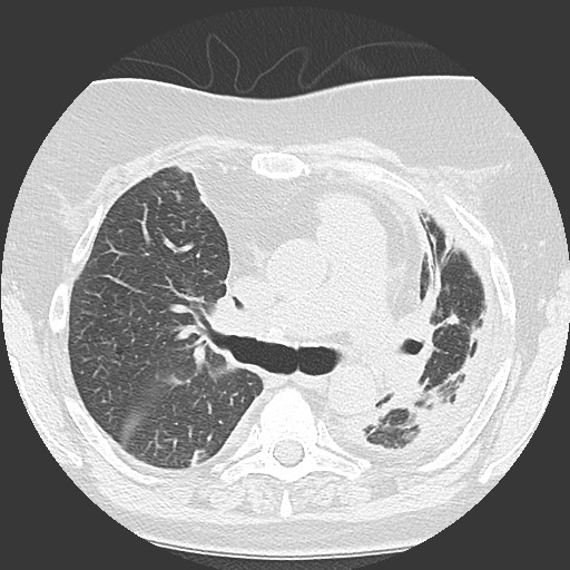
File:Chronic lung allograft dysfunction - restrictive form (Radiopaedia 60595-68316 Axial lung window 28).jpg
Jump to navigation
Jump to search
Chronic_lung_allograft_dysfunction_-_restrictive_form_(Radiopaedia_60595-68316_Axial_lung_window_28).jpg (512 × 512 pixels, file size: 75 KB, MIME type: image/jpeg)
Summary:
| Description |
|
| Date | Published: 8th Jun 2018 |
| Source | https://radiopaedia.org/cases/chronic-lung-allograft-dysfunction-restrictive-form |
| Author | Bruno Di Muzio |
| Permission (Permission-reusing-text) |
http://creativecommons.org/licenses/by-nc-sa/3.0/ |
Licensing:
Attribution-NonCommercial-ShareAlike 3.0 Unported (CC BY-NC-SA 3.0)
File history
Click on a date/time to view the file as it appeared at that time.
| Date/Time | Thumbnail | Dimensions | User | Comment | |
|---|---|---|---|---|---|
| current | 03:32, 22 August 2021 |  | 512 × 512 (75 KB) | Fæ (talk | contribs) | Radiopaedia project rID:60595 (batch #8055-47 B28) |
You cannot overwrite this file.
File usage
The following page uses this file:
