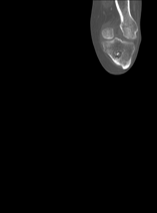
File:Chronic osteomyelitis - tibia (with sequestrum) (Radiopaedia 73273-84090 Coronal non-contrast 8).jpg
Jump to navigation
Jump to search
Size of this preview: 443 × 600 pixels. Other resolutions: 177 × 240 pixels | 627 × 849 pixels.
Original file (627 × 849 pixels, file size: 21 KB, MIME type: image/jpeg)
Summary:
| Description |
|
| Date | Published: 7th Jan 2020 |
| Source | https://radiopaedia.org/cases/chronic-osteomyelitis-tibia-with-sequestrum |
| Author | Utkarsh Kabra |
| Permission (Permission-reusing-text) |
http://creativecommons.org/licenses/by-nc-sa/3.0/ |
Licensing:
Attribution-NonCommercial-ShareAlike 3.0 Unported (CC BY-NC-SA 3.0)
File history
Click on a date/time to view the file as it appeared at that time.
| Date/Time | Thumbnail | Dimensions | User | Comment | |
|---|---|---|---|---|---|
| current | 18:07, 22 August 2021 | | 627 × 849 (21 KB) | Fæ (talk | contribs) | Radiopaedia project rID:73273 (batch #8103-8 A8) |
You cannot overwrite this file.
File usage
The following page uses this file:
_(Radiopaedia_73273-84090_Coronal_non-contrast_8).jpg&oldid=1082769)