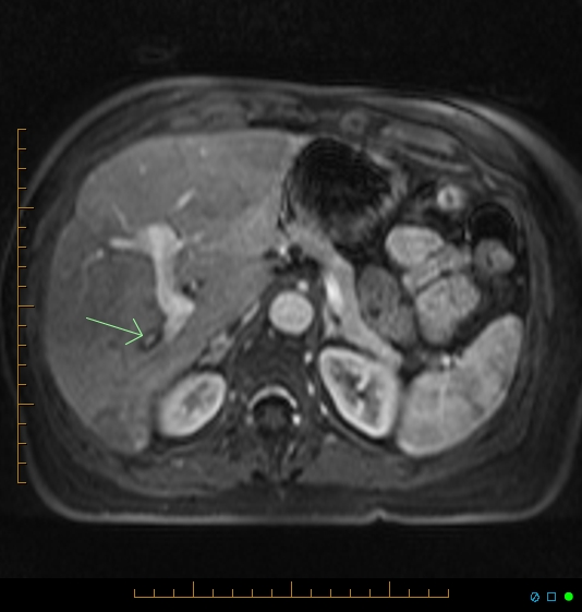
File:Chronic right portal vein occlusion (Radiopaedia 26946-27126 C 1).jpg
Jump to navigation
Jump to search
Chronic_right_portal_vein_occlusion_(Radiopaedia_26946-27126_C_1).jpg (534 × 561 pixels, file size: 102 KB, MIME type: image/jpeg)
Summary:
| Description |
|
| Date | Published: 15th Jan 2014 |
| Source | https://radiopaedia.org/cases/chronic-right-portal-vein-occlusion |
| Author | Chris O'Donnell |
| Permission (Permission-reusing-text) |
http://creativecommons.org/licenses/by-nc-sa/3.0/ |
Licensing:
Attribution-NonCommercial-ShareAlike 3.0 Unported (CC BY-NC-SA 3.0)
File history
Click on a date/time to view the file as it appeared at that time.
| Date/Time | Thumbnail | Dimensions | User | Comment | |
|---|---|---|---|---|---|
| current | 13:42, 23 August 2021 |  | 534 × 561 (102 KB) | Fæ (talk | contribs) | Radiopaedia project rID:26946 (batch #8158-3 C1) |
You cannot overwrite this file.
File usage
There are no pages that use this file.
