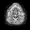
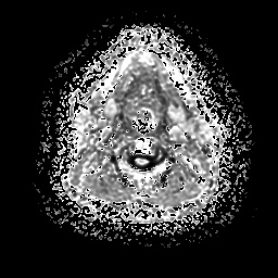
File:Chronic submandibular sialadenitis (Radiopaedia 61852-69885 Axial eADC 14).jpg
Jump to navigation
Jump to search
Chronic_submandibular_sialadenitis_(Radiopaedia_61852-69885_Axial_eADC_14).jpg (256 × 256 pixels, file size: 35 KB, MIME type: image/jpeg)
Summary:
| Description |
|
| Date | Published: 20th Jul 2018 |
| Source | https://radiopaedia.org/cases/chronic-submandibular-sialadenitis |
| Author | Varun Babu |
| Permission (Permission-reusing-text) |
http://creativecommons.org/licenses/by-nc-sa/3.0/ |
Licensing:
Attribution-NonCommercial-ShareAlike 3.0 Unported (CC BY-NC-SA 3.0)
File history
Click on a date/time to view the file as it appeared at that time.
| Date/Time | Thumbnail | Dimensions | User | Comment | |
|---|---|---|---|---|---|
| current | 20:26, 23 August 2021 |  | 256 × 256 (35 KB) | Fæ (talk | contribs) | Radiopaedia project rID:61852 (batch #8191-194 F14) |
You cannot overwrite this file.
File usage
There are no pages that use this file.
