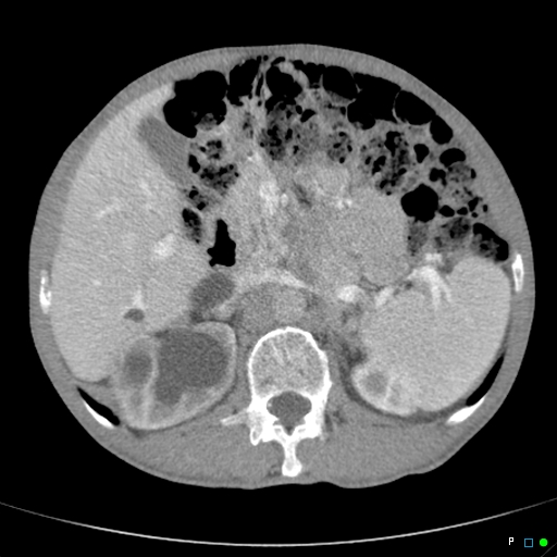
File:Chronic urinary retention (Radiopaedia 34678-36095 A 5).jpg
Jump to navigation
Jump to search
Chronic_urinary_retention_(Radiopaedia_34678-36095_A_5).jpg (512 × 512 pixels, file size: 112 KB, MIME type: image/jpeg)
Summary:
| Description |
|
| Date | Published: 4th Mar 2015 |
| Source | https://radiopaedia.org/cases/chronic-urinary-retention |
| Author | Chris O'Donnell |
| Permission (Permission-reusing-text) |
http://creativecommons.org/licenses/by-nc-sa/3.0/ |
Licensing:
Attribution-NonCommercial-ShareAlike 3.0 Unported (CC BY-NC-SA 3.0)
File history
Click on a date/time to view the file as it appeared at that time.
| Date/Time | Thumbnail | Dimensions | User | Comment | |
|---|---|---|---|---|---|
| current | 01:26, 24 August 2021 |  | 512 × 512 (112 KB) | Fæ (talk | contribs) | Radiopaedia project rID:34678 (batch #8205-5 A5) |
You cannot overwrite this file.
File usage
The following page uses this file:
