File:Chronic urinary retention (Radiopaedia 34678-36096 Axial C+ delayed 26).jpg

From NC Commons
Jump to navigation Jump to search

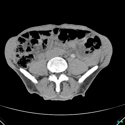
Chronic_urinary_retention_(Radiopaedia_34678-36096_Axial_C+_delayed_26).jpg(512 × 512 pixels, file size: 82 KB, MIME type: image/jpeg)

Summary:

Description
  • Radiopaedia case ID: 34678
  • Image ID: 11477718
  • Image stack position: 26/58
  • Plane projection: Axial
  • Aux modality: C+ delayed
  • Study description: Repeat CT following insertion of urinary catheter at 2100 hours
  • Study findings: Following catheter insertion and 5 hour delay the mass has "disappeared" with 4 liters of urine in the catheter bag. The bladder is collapsed and markedly thick-walled. Note contrast in the dilated left renal collecting system down to the bladder, but poorly functioning right kidney due to chronic backflow obstruction. Cause is presumed to be bladder outlet obstruction (possible due to the prostate or other urethral stricture). Note the so-called stasis bladder stone.
  • Modality: CT
  • System: Urogenital
  • Findings: Very large bilobed cystic mass arising in the pelvis with gross hydronephrosis and hydroureter right > left. Mass has 2 lobes: on the left is thick-walled and trabeculated (containing a calculus) consistent with the bladder and on the right is thin-walled and communicates with the bladder via a defect in the wall = pseudodiverticulum. Left leg swelling is due to extrinsic compression of the left iliac veins.
Date Published: 4th Mar 2015
Source https://radiopaedia.org/cases/chronic-urinary-retention
Author Chris O'Donnell
Permission
(Permission-reusing-text)
http://creativecommons.org/licenses/by-nc-sa/3.0/

Licensing:

Attribution-NonCommercial-ShareAlike 3.0 Unported (CC BY-NC-SA 3.0)

File history

Click on a date/time to view the file as it appeared at that time.

Date/TimeThumbnailDimensionsUserComment
current01:45, 24 August 2021Thumbnail for version as of 01:45, 24 August 2021512 × 512 (82 KB) (talk | contribs)Radiopaedia project rID:34678 (batch #8205-26 A26)