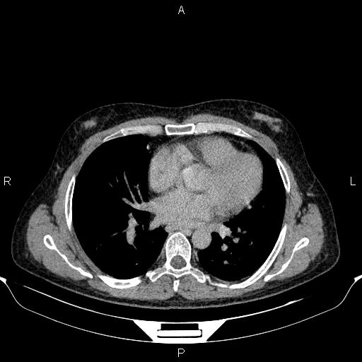
File:Cirrhosis (Radiopaedia 86990-103221 Axial C+ delayed 1).jpg
Jump to navigation
Jump to search
Cirrhosis_(Radiopaedia_86990-103221_Axial_C+_delayed_1).jpg (512 × 512 pixels, file size: 31 KB, MIME type: image/jpeg)
Summary:
| Description |
|
| Date | Published: 18th Feb 2021 |
| Source | https://radiopaedia.org/cases/cirrhosis-17 |
| Author | Mohammad Taghi Niknejad |
| Permission (Permission-reusing-text) |
http://creativecommons.org/licenses/by-nc-sa/3.0/ |
Licensing:
Attribution-NonCommercial-ShareAlike 3.0 Unported (CC BY-NC-SA 3.0)
File history
Click on a date/time to view the file as it appeared at that time.
| Date/Time | Thumbnail | Dimensions | User | Comment | |
|---|---|---|---|---|---|
| current | 12:39, 24 August 2021 |  | 512 × 512 (31 KB) | Fæ (talk | contribs) | Radiopaedia project rID:86990 (batch #8235-147 C1) |
You cannot overwrite this file.
File usage
The following page uses this file:
