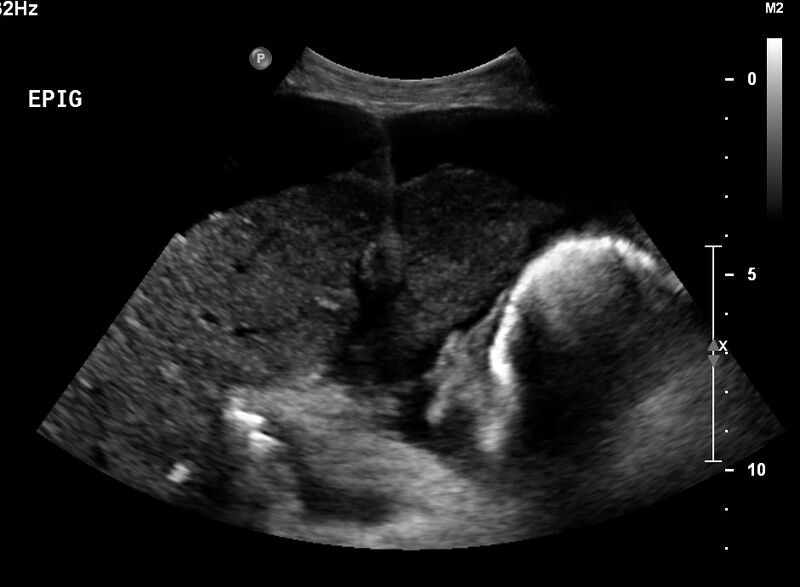
File:Cirrhosis and portal hypertension (Radiopaedia 79127).jpg
Jump to navigation
Jump to search

Size of this preview: 800 × 587 pixels. Other resolutions: 320 × 235 pixels | 640 × 470 pixels | 941 × 691 pixels.
Original file (941 × 691 pixels, file size: 62 KB, MIME type: image/jpeg)
Summary:
| Description |
|
| Date | Published: 21st Jun 2020 |
| Source | https://radiopaedia.org/cases/cirrhosis-and-portal-hypertension-1 |
| Author | Bruno Di Muzio |
| Permission (Permission-reusing-text) |
http://creativecommons.org/licenses/by-nc-sa/3.0/ |
Licensing:
Attribution-NonCommercial-ShareAlike 3.0 Unported (CC BY-NC-SA 3.0)
File history
Click on a date/time to view the file as it appeared at that time.
| Date/Time | Thumbnail | Dimensions | User | Comment | |
|---|---|---|---|---|---|
| current | 22:06, 24 August 2021 | | 941 × 691 (62 KB) | Fæ (talk | contribs) | Radiopaedia project rID:79127 (batch #8250) |
You cannot overwrite this file.
File usage
The following page uses this file:
.jpg&oldid=8852858)