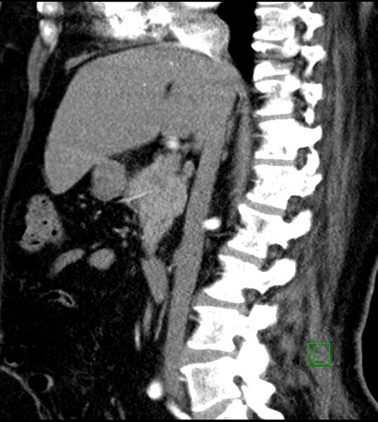
File:Clear cell renal cell carcinoma (Radiopaedia 85004-100537 D 35).jpg

From NC Commons
Jump to navigation Jump to search

Original file(628 × 701 pixels, file size: 98 KB, MIME type: image/jpeg)

Summary:

Description
  • Radiopaedia case ID: 85004
  • Image ID: 54046269
  • Image stack position: 35/61
  • Plane projection: Sagittal
  • Aux modality: C+ arterial phase
  • Study findings: Findings: Large lobulated exophytic mass lesion measuring 10 x 8.8 x 8.5 cm is seen at the lower pole of the right kidney. Two tiny calcification foci are seen in it. It has an enhancing (non-contrast density=20 HU, Venous phase density=82 HU) peripheral solid component and non-enhancing irregular necrotic center. The mass is grossly extending beyond the renal capsule, has mild perinephric fat stranding. However, it is still confined within Gerota's fascia. Some prominent arteries & veins (neo-vascularity) are seen along the inferior aspect of this mass lesion. A prominent draining vein is seen at the inferior aspect of this mass, which is draining to the inferior vena cava via the right ovarian vein. The mass is compressing the lower pole calyceal system and the renal pelvis; however, no invasion/infiltration of the collecting system or hydronephrosis is seen. No clear signs of the adjacent renal vein or IVC thrombosis. No evidence of local or distant metastasis is seen. A small simple cyst is seen in either kidney.  Multiple focal radiolucent lesions with polka dot sign in the axial images are seen in the T11, T12, L1, and L2 vertebral bodies; these are likely vertebral hemangiomas. Impression: Large heterogeneous right renal mass, suspicious of renal cell carcinoma. Note: This is an old case. Unfortunately the "axial venous phase images" are not available.
  • Modality: CT
  • System: Oncology
  • Findings: A large exophytic lobulated heterogeneous solid mass lesion measuring 7.9 x 8.4 cm is depicted along the mid/lower pole of the right kidney. Small fluid echogenicity areas, likely representing necrosis, are seen within the center of this lesion. Some vascularity is seen in its peripheral part on Doppler ultrasound examination. Impression: Highly suspicious right renal mass lesion.
Date Published: 12th Dec 2020
Source https://radiopaedia.org/cases/clear-cell-renal-cell-carcinoma-7
Author Ammar Ashraf
Permission
(Permission-reusing-text)
http://creativecommons.org/licenses/by-nc-sa/3.0/

Licensing:

Attribution-NonCommercial-ShareAlike 3.0 Unported (CC BY-NC-SA 3.0)

File history

Click on a date/time to view the file as it appeared at that time.

Date/TimeThumbnailDimensionsUserComment
current17:13, 25 August 2021Thumbnail for version as of 17:13, 25 August 2021628 × 701 (98 KB) (talk | contribs)Radiopaedia project rID:85004 (batch #8323-172 D35)