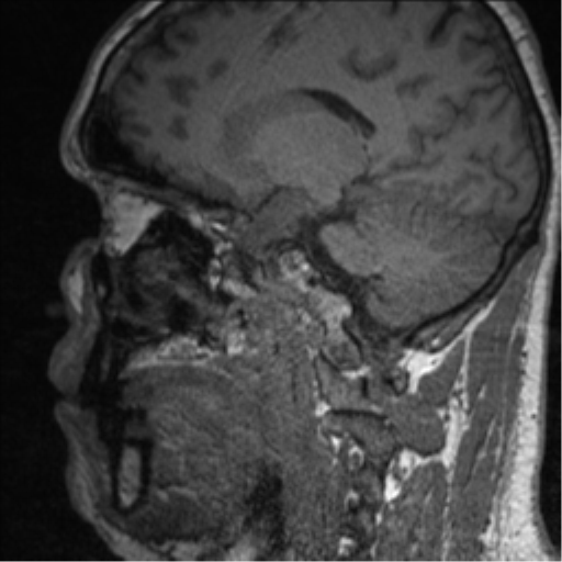
File:Clival chordoma invading the hypoglossal canal (Radiopaedia 48850-53884 Sagittal T1 45).png
Jump to navigation
Jump to search
No higher resolution available.
Clival_chordoma_invading_the_hypoglossal_canal_(Radiopaedia_48850-53884_Sagittal_T1_45).png (512 × 512 pixels, file size: 123 KB, MIME type: image/png)
Summary:
| Description |
|
| Date | Published: 1st Nov 2016 |
| Source | https://radiopaedia.org/cases/clival-chordoma-invading-the-hypoglossal-canal |
| Author | Bruno Di Muzio |
| Permission (Permission-reusing-text) |
http://creativecommons.org/licenses/by-nc-sa/3.0/ |
Licensing:
Attribution-NonCommercial-ShareAlike 3.0 Unported (CC BY-NC-SA 3.0)
File history
Click on a date/time to view the file as it appeared at that time.
| Date/Time | Thumbnail | Dimensions | User | Comment | |
|---|---|---|---|---|---|
| current | 05:12, 26 August 2021 | | 512 × 512 (123 KB) | Fæ (talk | contribs) | Radiopaedia project rID:48850 (batch #8380-137 E45) |
You cannot overwrite this file.
File usage
The following page uses this file:
.png&oldid=1132887)