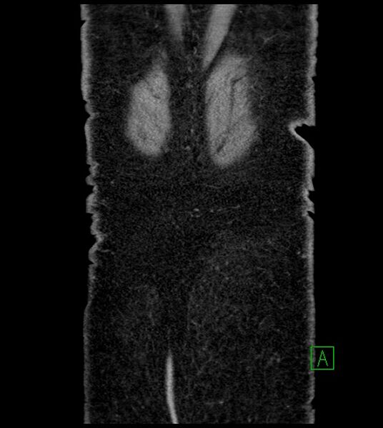
File:Closed-loop small bowel obstruction-internal hernia (Radiopaedia 78439-91096 Coronal non-contrast 100).jpg

From NC Commons
Jump to navigation Jump to search

Original file(630 × 705 pixels, file size: 74 KB, MIME type: image/jpeg)

Summary:

Description
  • Radiopaedia case ID: 78439
  • Image ID: 52813996
  • Image stack position: 100/109
  • Plane projection: Coronal
  • Aux modality: non-contrast
  • Study findings: Changes of previous biliopancreatic diversion surgery are seen. Dilated oral contrast filled jejunal loops measuring 3-4 cm are seen in the left lumbar region. No oral contrast passage is seen in the distal small bowel. Mildly dilated fluid-filled ileal loops are seen clustered in the right lumbar region. Two distinct transition points located next to each other, mesenteric edema/inter-loop fluid, and a whirl sign are appreciable along these dilated ileal loops in the right lumbar region.  No pneumatosis or pneumoperitoneum is seen. A small amount of free fluid is seen around the liver. No oral contrast leakage/extravasation is seen. A hyperdense focus is seen in supra-renal abdominal inferior vena cava (inferior vena cava filter with probable surrounding thrombus). Enlarged azygous vein and multiple collaterals are seen in the abdominal cavity as well as in the subcutaneous soft tissues of the abdominal wall; these findings are suggestive of chronic inferior vena cava thrombosis (likely as a complication of IVC filter or a complication of previous bariatric surgery). Average size kidneys with multiple peripheral small subcentimeter hyperdense cysts (average density=70-90 HU), which are likely hemorrhagic cysts (hyperdense renal cysts). A simple cortical cyst is also seen in either kidney. CONCLUSION: The above mentioned CT findings are suggestive of closed-loop small bowel obstruction secondary to internal hernia (related to previous surgical interventions); another possibility can be adhesions. The possibility of small bowel ischemia cannot be excluded from this unenhanced examination (IV contrast not administered due to high creatinine).
  • Modality: CT
  • System: Gastrointestinal
  • Findings: A few mildly dilated small bowel loops with multiple air fluid levels are seen. No pneumoperitoneum is seen.  A few surgical clips are seen in the epigastrium and right hypochondrium.
Date Published: 4th Jun 2020
Source https://radiopaedia.org/cases/closed-loop-small-bowel-obstruction-internal-hernia-3
Author Ammar Ashraf
Permission
(Permission-reusing-text)
http://creativecommons.org/licenses/by-nc-sa/3.0/

Licensing:

Attribution-NonCommercial-ShareAlike 3.0 Unported (CC BY-NC-SA 3.0)

File history

Click on a date/time to view the file as it appeared at that time.

Date/TimeThumbnailDimensionsUserComment
current18:31, 27 August 2021Thumbnail for version as of 18:31, 27 August 2021630 × 705 (74 KB) (talk | contribs)Radiopaedia project rID:78439 (batch #8425-265 B100)