File:Closed loop obstruction (Radiopaedia 33319-34356 A 25).png

From NC Commons
Jump to navigation Jump to search

Closed_loop_obstruction_(Radiopaedia_33319-34356_A_25).png(512 × 512 pixels, file size: 161 KB, MIME type: image/png)

Summary:

Description
  • Radiopaedia case ID: 33319
  • Image ID: 10235065
  • Image stack position: 25/78
  • Plane projection: AXIAL THICK
  • Aux modality: C+ portal venous phase
  • Modality: CT
  • System: Gastrointestinal
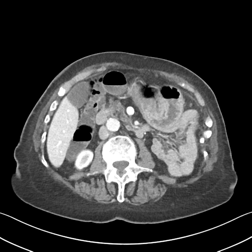
  • Findings: Previous colectomy. The oversewn rectosigmoid is pulled upwards and to the right. The suture site is unremarkable. Submucosal fat within this remnant large bowel is in keeping with chronic inflammation secondary to the known history of IBD. Left iliac fossa end ileostomy, and associated parastomal hernia. At the neck of the hernia, three adjacent small bowel transition points are identified on a single axial slice. There is a closed loop obstruction of a small loop of small bowel that lies within the parastomal hernial sac. This loop of bowel is mildly distended and thick walled. The associated mesentery within the hernial sac demonstrates venous congestion, and there is a small pocket of free fluid within the hernial sac. There is a second closed loop obstruction of small bowel that lies within the peritoneal cavity, the point of obstruction also being at the level of the hernial neck. The small bowel involved is dilated up to 57mm. Upstream of these closed loop obstructions, there is a small left inguinal hernia that contains a loop of unremarkable small bowel. No pneumatosis intestinalis identified. The proximal jejunum is collapsed. No focal liver lesion. Indentation of segment 6 by a prominent diaphragmatic slip is noted. The gallbladder, portal vein, spleen, adrenal glands and kidneys have an unremarkable appearance other than a 15 mm interpolar renal cyst on the left. Other than the aforementioned free fluid within the parastomal hernial sac, no free intraperitoneal fluid identified. There is an irregular cystic lesion in the tail of pancreas with associated focus of internal calcification that measures 24mm. Within the uncinate process, there is a 29 mm cystic lesion. There is also a smaller rounded cystic lesion in the inferior portion of the head of pancreas that measures approximately 9 mm. The main pancreatic duct at the level of the pancreatic head measures 5 mm and is stable. The common bile duct is not dilated. Old left superior and inferior pubic rami fractures. Multiple vertebral compression fractures appear chronic. Conclusion: Left iliac fossa parastomal hernia, with two separate closed loop small bowel obstructions, one within the parastomal hernial sac, the other within the peritoneal cavity. Uncomplicated left inguinal hernia. Multiple cystic lesions within the tail and uncinate process of the pancreas.
Date Published: 7th Jan 2015
Source https://radiopaedia.org/cases/closed-loop-obstruction-2
Author Kenny Sim
Permission
(Permission-reusing-text)
http://creativecommons.org/licenses/by-nc-sa/3.0/

Licensing:

Attribution-NonCommercial-ShareAlike 3.0 Unported (CC BY-NC-SA 3.0)

File history

Click on a date/time to view the file as it appeared at that time.

Date/TimeThumbnailDimensionsUserComment
current13:54, 26 August 2021Thumbnail for version as of 13:54, 26 August 2021512 × 512 (161 KB) (talk | contribs)Radiopaedia project rID:33319 (batch #8395-25 A25)

The following page uses this file:

Metadata