File:Closed loop obstruction (Radiopaedia 86962-103314 Axial C+ 26).jpg

From NC Commons
Jump to navigation Jump to search

Closed_loop_obstruction_(Radiopaedia_86962-103314_Axial_C+_26).jpg(432 × 350 pixels, file size: 29 KB, MIME type: image/jpeg)

Summary:

Description
  • Radiopaedia case ID: 86962
  • Image ID: 54448448
  • Image stack position: 26/70
  • Plane projection: Axial C+
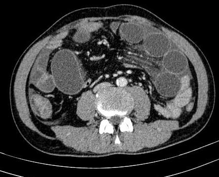
  • Study findings: CT findings demonstrate the typical features of closed-loop obstruction in SBO. Pink arrow: proximal/afferent loop Green arrow: closed-loop (strangulated loop) Blue arrow: distal/efferent loop Red circles: Two-point of obstruction Orange arrows: a clump of bowel loops Purple star:  mesenteric edema
  • Modality: Annotated image
  • System: Gastrointestinal
  • Findings: CT findings show dilated loops of the small bowel with gas-fluid levels, the transitions are at two adjacent points along the course of dilated bowel (double beak sign at two sites of fusiform tapering). The bowel loops have a strange arrangement: The clump of dilated loops concentrates at the periumbilical region with the mesenteric vessels towards a central point. The wall bowel has a normal enhancement but mesenteric edema was suggestive of ischemic bowel. The whirl sign can be seen on coronal reconstruction. Intraperitoneal free fluid in the recto-vesical pouch, the left iliac fossa, and surrounding the liver.
Date Published: 20th Feb 2021
Source https://radiopaedia.org/cases/closed-loop-obstruction-9
Author Luu Hanh
Permission
(Permission-reusing-text)
http://creativecommons.org/licenses/by-nc-sa/3.0/

Licensing:

Attribution-NonCommercial-ShareAlike 3.0 Unported (CC BY-NC-SA 3.0)

File history

Click on a date/time to view the file as it appeared at that time.

Date/TimeThumbnailDimensionsUserComment
current16:22, 26 August 2021Thumbnail for version as of 16:22, 26 August 2021432 × 350 (29 KB) (talk | contribs)Radiopaedia project rID:86962 (batch #8397-26 A26)

The following file is a duplicate of this file (more details):