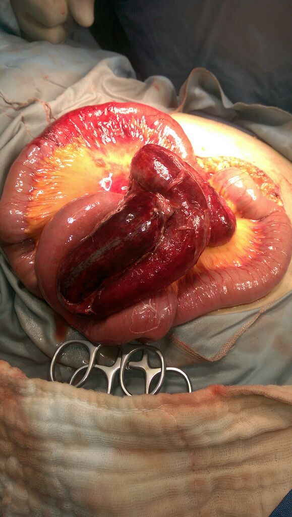
File:Closed loop small bowel obstruction (Radiopaedia 42193-45255 A 1).jpg
Jump to navigation
Jump to search

Size of this preview: 339 × 599 pixels. Other resolutions: 135 × 240 pixels | 271 × 480 pixels | 434 × 768 pixels | 579 × 1,024 pixels | 1,520 × 2,688 pixels.
Original file (1,520 × 2,688 pixels, file size: 1.31 MB, MIME type: image/jpeg)
Summary:
| Description |
|
| Date | Published: 10th Jan 2016 |
| Source | https://radiopaedia.org/cases/closed-loop-small-bowel-obstruction-6 |
| Author | Sakher Alkhaderi |
| Permission (Permission-reusing-text) |
http://creativecommons.org/licenses/by-nc-sa/3.0/ |
Licensing:
Attribution-NonCommercial-ShareAlike 3.0 Unported (CC BY-NC-SA 3.0)
File history
Click on a date/time to view the file as it appeared at that time.
| Date/Time | Thumbnail | Dimensions | User | Comment | |
|---|---|---|---|---|---|
| current | 01:25, 27 August 2021 | | 1,520 × 2,688 (1.31 MB) | Fæ (talk | contribs) | Radiopaedia project rID:42193 (batch #8409-1 A1) |
You cannot overwrite this file.
File usage
There are no pages that use this file.
.jpg&oldid=1147230)