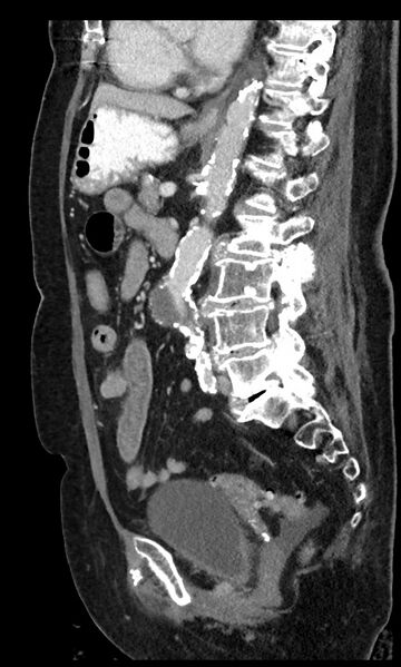
File:Closed loop small bowel obstruction - adhesions (Radiopaedia 59269-66611 C 51).jpg
Jump to navigation
Jump to search

Size of this preview: 360 × 599 pixels. Other resolutions: 144 × 240 pixels | 512 × 852 pixels.
Original file (512 × 852 pixels, file size: 106 KB, MIME type: image/jpeg)
Summary:
| Description |
|
| Date | 30 Mar 2018 |
| Source | Closed loop small bowel obstruction - adhesions |
| Author | Michael P Hartung |
| Permission (Permission-reusing-text) |
http://creativecommons.org/licenses/by-nc-sa/3.0/ |
Licensing:
Attribution-NonCommercial-ShareAlike 3.0 Unported (CC BY-NC-SA 3.0)
| This file is ineligible for copyright and therefore in the public domain, because it is a technical image created as part of a standard medical diagnostic procedure. No creative element rising above the threshold of originality was involved in its production.
|
File history
Click on a date/time to view the file as it appeared at that time.
| Date/Time | Thumbnail | Dimensions | User | Comment | |
|---|---|---|---|---|---|
| current | 05:16, 27 August 2021 |  | 512 × 852 (106 KB) | Fæ (talk | contribs) | Radiopaedia project rID:59269 (batch #8413-332 C51) |
You cannot overwrite this file.
File usage
The following page uses this file:
