File:Closed loop small bowel obstruction in pregnancy (MRI) (Radiopaedia 87637-104031 D 39).jpg
Jump to navigation
Jump to search
No higher resolution available.
Closed_loop_small_bowel_obstruction_in_pregnancy_(MRI)_(Radiopaedia_87637-104031_D_39).jpg (256 × 192 pixels, file size: 19 KB, MIME type: image/jpeg)
Summary:
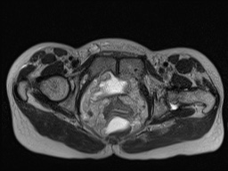
| Description |
|
| Date | Published: 11th Mar 2021 |
| Source | https://radiopaedia.org/cases/closed-loop-small-bowel-obstruction-in-pregnancy-mri |
| Author | Andrew Plumb |
| Permission (Permission-reusing-text) |
http://creativecommons.org/licenses/by-nc-sa/3.0/ |
Licensing:
Attribution-NonCommercial-ShareAlike 3.0 Unported (CC BY-NC-SA 3.0)
File history
Click on a date/time to view the file as it appeared at that time.
| Date/Time | Thumbnail | Dimensions | User | Comment | |
|---|---|---|---|---|---|
| current | 15:40, 27 August 2021 | | 256 × 192 (19 KB) | Fæ (talk | contribs) | Radiopaedia project rID:87637 (batch #8422-184 D39) |
You cannot overwrite this file.
File usage
The following page uses this file:
_(Radiopaedia_87637-104031_D_39).jpg&oldid=1157525)